
 |
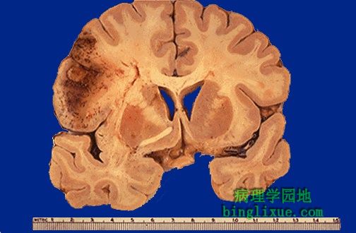
动脉栓子引起的脑梗死,常导致出血性外观。水肿使结构模糊不清。急性水肿性梗死组织可形成团块效应。可见左侧正中线移位,脑室缩小。 Here is a cerebral infarct from an arterial embolus, which often leads to a hemorrhagic appearance. There is edema which obscures the structures. The acutely edematous infarcted tissue may produce a mass effect. Note the decrease in size of the ventricle on the left with shift of the midline. |
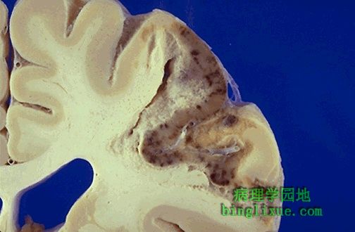 |
图示梗死为小斑点状出血,梗死由栓子引起。 The infarction seen here has punctate hemorrhages. This infarct was caused by an embolus. |
 |
急性脑梗死的显微镜图像显示明显水肿(苍白色区域)。 The microscopic appearance of this acute cerebral infarction reveals marked edema (the pale areas). |
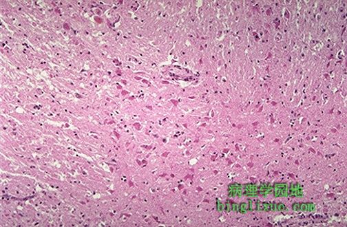 |
神经元是对缺氧最敏感的细胞。图中可见的红色神经元死于缺氧。 The neurons are the most sensitive cells to anoxic injury. Seen here are red neurons which are dying as a result of hypoxia. |
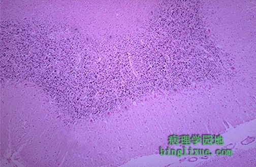 |
小脑分子层和颗粒层之间的浦肯野细胞对缺氧也是非常敏感的。图示浦肯野细胞是红色的。 The Purkinje cells between the molecular and granular layers of the cerebellum are also highly susceptible to anoxia. Those seen here are red. |
 |
右边是亚急性梗死(中速),出现明显水肿,伴组织结构轮廓不清和肿胀,正中线推向左侧。发生液化性坏死并形成囊性空腔。 The subacute (intermediate) infarct seen here at the right shows edema with obscured structural outlines and swelling that shifts the midline to the left. There is liquefactive necrosis with beginning formation of cystic spaces. |
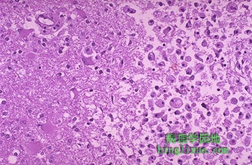 |
脑梗死显微镜外观,右侧出现众多巨噬细胞,它们正清理液化性坏死造成的脂质碎片。 This cerebral infarction demonstrates the presence of many macrophages at the right which are cleaning up the lipid debris from the liquefactive necrosis. |
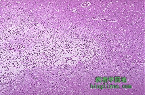 |
脑梗死灶中的液化性坏死的溶解导致囊性空腔形成。 Resolution of the liquefactive necrosis in a cerebral infarction leads to the formation of a cystic space. |
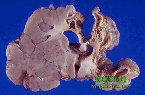 |
新生儿大的陈旧性脑梗死灶。液化性坏死的溶解留下了一个巨大的囊性空腔,它包绕大脑半球的大部分。 Here is a large remote cerebral infarction. Resolution of the infarction has left a huge cystic space encompassing much of the cerebral hemisphere in this neonate. |
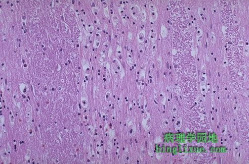 |
脑干高倍镜显示,脑梗死可同时伴有下行束沃勒变性。 Cerebral infarctions can be accompanied by Wallerian degeneration of descending tracts, as shown here at high power in the brainstem. |