
 |
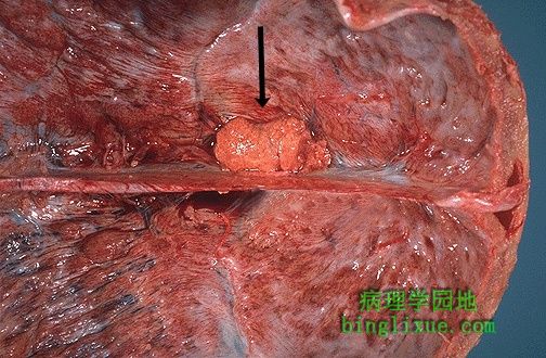
边界清晰、红黄的质地坚韧的脑膜瘤,位于硬脑膜下,临近大脑镰。发生于正中线旁且偏上的部位是相当普通的。 This circumscribed reddish-yellow firm neoplasm beneath the dura next to the falx is a meningioma. The superior parasagittal location is quite common. |
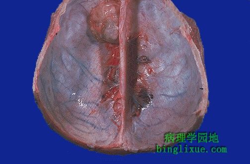 |
硬脑膜下另例良性脑膜瘤,生长缓慢,但症状被发现前,能长的很大。 Here is another benign meningioma beneath the dura. These neoplasms are slow growing, but may reach a large size before symptoms lead to detection. |
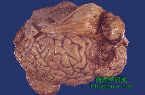 |
可见脑膜下的脑膜瘤是如何压迫下面的大脑半球的。浸润性脑膜瘤罕见。 Note how this meningioma beneath the dura has compressed the underlying cerebral hemisphere. Rarely, meningiomas can be more aggressive and invade. |
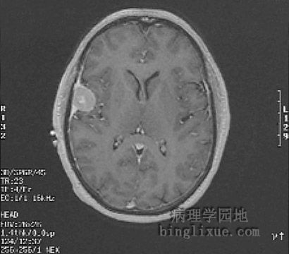 |
MRI清晰可见右侧一肿块压迫大脑半球。病变特征与脑膜瘤一致。 This is an MRI scan demonstrating a discreet mass along the lateral convexity and extending from a dural base impinging upon the cerebral hemisphere. This is consistent with a meningioma. |
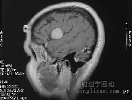 |
矢状面MRI显示脑膜瘤。 A sagittal view is shown here. |
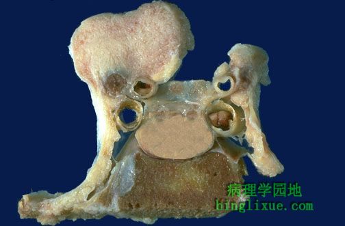 |
尸检中偶然发现的位于蝶骨脊处的脑膜瘤(左上),它是脑膜瘤的另一个好发部位。蝶鞍底位于中间下部,它的上面是灰褐色垂体。垂体的上部及侧部是颈内动脉和大脑前动脉,再稍微向上即是位于中间的视神经(略带黄色)。 An incidental finding at autopsy, this light tan colored meningioma involved the sphenoid ridge, another common location. The base of the sella is seen here, above which is the tan-colored pituitary gland. Above and on each side lateral to pituitary are the internal carotid and the anterior cerebral arteries, above which slightly medially are the optic nerves. |
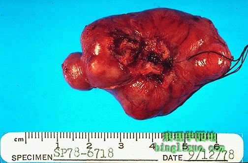 |
已切除的脑膜瘤。对于神经外科医生来说,切除这类肿瘤相当容易。 Here is a resected meningioma. These are relatively easy for the neurosurgeon to remove. |
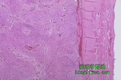 |
低倍镜脑膜瘤图像。右边是致密的粉红色结缔组织硬脑膜。脑膜瘤细胞有大量的粉红色胞质。 This is the microscopic appearance of a meningioma of a meningioma at low magnification. Note the dense pink connective tissue dura at the right. The cells of the meningioma have abundant pink cytoplasm. |
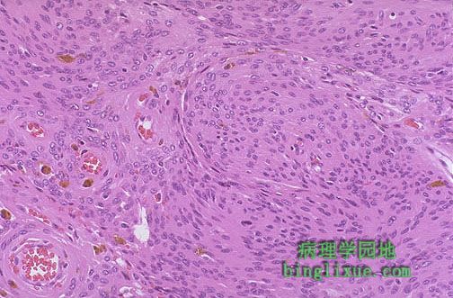 |
中倍镜,脑膜瘤由涡漩状细胞巢构成,也可能有多种形状。 At medium power, this meningioma is composed of whorled nests of cells. A variety of patterns are possible. |
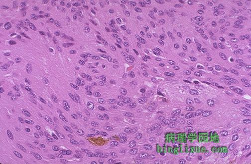 |
高倍镜,脑膜瘤中存在饱满的粉红色细胞。可见少量棕色颗粒状含铁血黃素。脑膜瘤也可以有砂粒体。 At high magnification, this meningioma has plump pink cells. A small amount of brown granular hemosiderin is present. Meningiomas may also have psammomma bodies. |