
 |
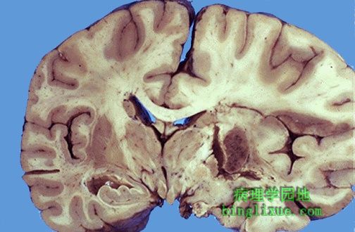
缓进型高血压小动脉硬化导致小范围的腔隙性梗死灶,脑桥上可见一此种病变。此病变多位于基底节、白质深部、脑干。 The arteriolar sclerosis that results from chronic hypertension leads to small lacunar infarcts, or "lacunes", one of which is seen here in the pons. Such lesions are most common in basal ganglia, deep white matter, and brain stem. |
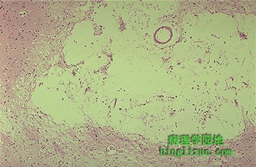 |
腔隙性梗死显微镜图像。注意液化性坏死溶解形成的囊性空腔。也可出现出血形成的含铁血黄素。 This is the microscopic appearance of a lacunar infarct. Note that it is a cystic space from the resolved liquefactive necrosis. There can be hemosiderin pigment from hemorrhage as well. |
 |
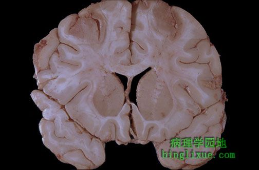
急性脑梗死,这种梗死是动脉血栓形成或栓塞的典型表现。 An acute cerebral infarct is seen here. Such infarcts are typically the result of arterial thrombosis or embolism. |
 |
明显双侧对称的黑的褪色区域,表示在大脑动脉环的前部和中部之间的区域最近出现的梗死。相对或绝对的脑低灌注都可能造成这种梗死。 The bilaterally symmetric dark discolored areas seen superiorly and just lateral to the midline represent recent infarction in the watershed zone between anterior and middle cerebral arterial circulations. Such watershed infarctions can occur with relative or absolute hypoperfusion of the brain. |
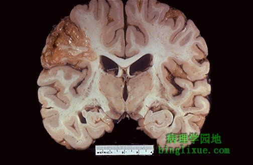 |
大脑中动脉供血区域的陈旧性梗死。 This is an intermediate to remote infarct in the distribution of the middle cerebral artery. |
 |
颈内动脉血栓形成。在脑部,动脉血栓比静脉血栓更常见(比例大约是100比1)。 A thrombosis of the internal carotid artery is seen here. Arterial thromboses are far more common in the brain than venous thromboses (by a ratio of about 100 to 1). |
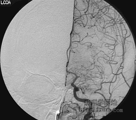 |
血管造影显示左颈总动脉的主要分支的栓子阻塞。 This angiogram demonstrates an embolic obstruction of a branch of the left common carotid artery just past the first main bifurcation. |
 |
血栓栓子可以栓塞于脑动脉,尤其是在大脑中动脉及其分支。此栓子源于左心房的附壁血栓。心脏是这种拴子的常见来源。 Thromboemboli can lodge in cerebral arteries, particularly in the distribution of the middle cerebral, and peripherally toward branch points. Here is a thromboembolus that originated from mural thrombus in the left atrium. The heart is a common source for such emboli. |
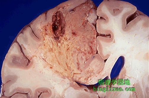 |
随着溶解开始,额叶亚急性梗死导致液化性坏死并伴有囊性空腔形成。 This intermediate infarct of the frontal lobe shows liquefactive necrosis with formation of cystic spaces as resolution begins. |
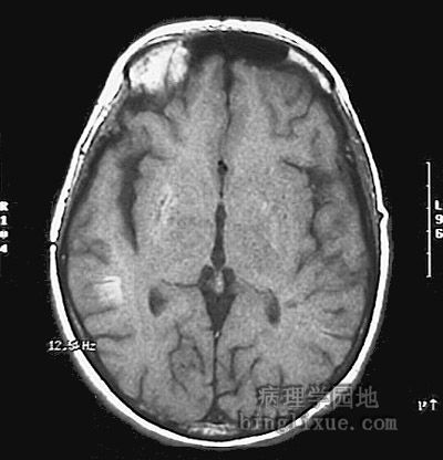 |
MRI显示右侧基底节和顶区后部的灰质白质交界处亚急性梗死。 This magnetic resonance imaging scan demonstrates subacute infarctions in the right basal ganglia and also near the gray-white junction in the posterior parietal region. |