
 |
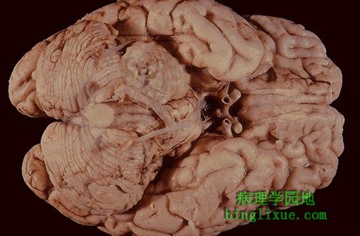
小脑桥脑角听神经(第八对脑神经)肿块病变,为小脑桥脑角神经鞘瘤,病人可出现听力降低。这类良性肿瘤可切除。 The mass lesion here is arising in the acoustic (eighth cranial) nerve at the cerebellopontine angle. This is a schwannoma. Patients may present with hearing loss. These benign neoplasms can be removed. |
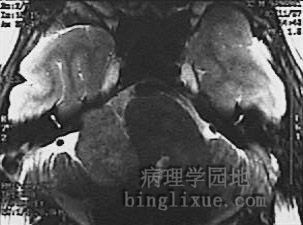 |
横断面MRI显示小脑桥脑角肿块压迫小脑,为神经鞘瘤,即听神经瘤(因累及Ⅷ脑神经)。 This magnetic resonance imaging (MRI) scan in transverse view demonstrates a mass impinging upon the cerebellum from the cerebellopontine angle. This is a schwannoma, also known as an acoustic neuroma because it arises from the eighth nerve. |
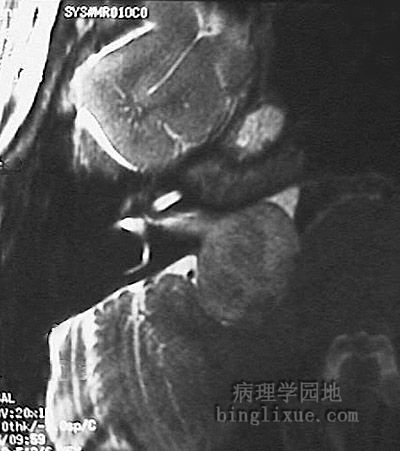 |
听神经瘤MRI放大图像。 Here is a magnified view of the acoustic neuroma. |
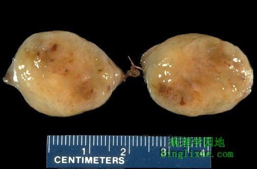 |
神经鞘瘤切面和许多间叶细胞肿瘤相似,具有鱼肉状、质地软、棕褐色外观。 The cut surface of a schwannoma is similar to that of many mesenchymal neoplasms, with a "fish flesh" soft tan appearance. |
 |
显微镜显示神经鞘瘤的典型结构,为良性肿瘤。注意左侧为多细胞的束状型(Antoni A 型),存在栅栏状细胞包围的粉红色区域(Verocay小体)。右边是网状型(Antoni B 型),间质疏松、细胞少、粘液样改变。 These are the classic microscopic appearances of a schwannoma, which is benign. Note the more cellular "Antoni A" pattern on the left with palisading nuclei surrounding pink areas (Verocay bodies). On the right is the "Antoni B" pattern with a looser stroma, fewer cells, and myxoid change. |
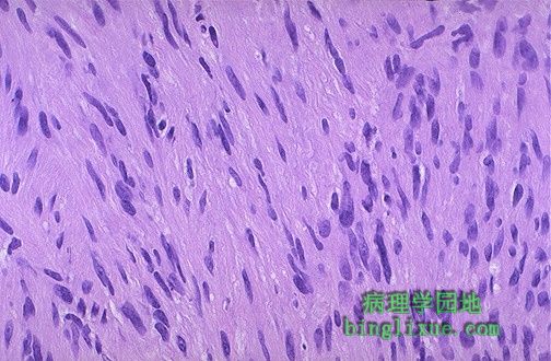 |
高倍镜显示神经鞘瘤。 The schwannoma is seen here at higher magnification. |
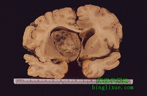 |
大脑半球胶质瘤显示的肿块病变。注意肿瘤颜色混杂,存在红色、褐色、白色以及棕色。 This glioma of the cerebral hemisphere demonstrates a mass effect. Note the variegation of the neoplasm, with areas of red, tan, white, and brown. |
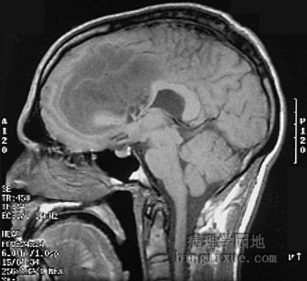 |
矢状面MRI显示大的胶质瘤压迫脑室。 This is a sagittal MRI scan demonstrating a large glioma impinging upon the ventricular system. |
 |
水平扫描MRI显示胶质瘤。 A transverse view is shown here. |
 |
即使胶质瘤分级较低,也仍然是恶性的,恶性程度与发生部位有关,脑干的胶质瘤最糟糕--不能被切除。 Even if a glioma is "low grade" it can still be bad, depending upon the location. This glioma of the brainstem is in the worst possible location-- it cannot be resected. |