
 |
链球菌感染后肾小球肾炎是由免疫介导的。免疫沉积物呈颗粒状分布于肾小球基底膜和系膜区。 Post-streptococcal glomerulonephritis is immunologically mediated, and the immune deposits are distributed in the capillary loops in a granular, bumpy pattern because of the focal nature of the deposition process. |
 |
电镜下,链球菌感染后肾小球肾炎的电子致密的免疫沉积物主要位于脏层上皮下,图示基底膜右侧一个大的脏层上皮下驼峰状沉积物。毛细血管腔内充满了中性粒细胞,它们的核呈分叶状,还可见细胞浆内的颗粒。 By electron microscopy, the electron dense immune deposits of post-streptococcal glomerulonephritis are predominantly subepithelial, as seen here with a large subepithelial "hump" at the right of the basement membrane (BM). The capillary lumen is filled with a PMN whose nuclear lobes and cytoplasmic granules are visible. |
 |
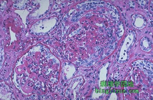
膜性肾小球肾炎,毛细血管袢明显增厚,但细胞并不增多。膜性肾小球肾炎是成年人肾病综合征最常见的原因。有些膜性肾小球肾炎和慢性感染(如乙型肝炎)、肿瘤或SLE有关。但多数病例是特发性的。 Here is the light microscopic appearance of membranous glomerulonephritis in which the capillary loops are thickened and prominent, but the cellularity is not increased. Membranous GN is the most common cause for nephrotic syndrome in adults. Some cases of membranous GN can be linked to a chronic infectious disease such as hepatitis B, a carcinoma, or SLE, but many cases are idiopathic. |
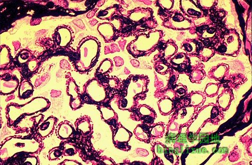 |
膜性肾小球肾炎,肾小球的银染显示呈黑色的基底膜及与之垂直排列的钉突。 A silver stain of the glomerulus highlights the proteinaceous basement membranes in black. There are characteristic "spikes" seen with membranous glomerulonephritis seen here in which the black basement membrane material appears as projections around the capillary loops. |
 |
膜性肾小球肾炎是一种免疫介导的疾病。如图所见:IgG和补体沉积于基底膜,表现为典型的颗粒状荧光。 Membranous glomerulonephritis is an immunologically mediated disease in which deposts of mainly IgG and complement collect in the basement membrane and appear in a diffuse granular pattern by immunofluorescence, as seen here. |
 |
电镜下,可见膜性肾小球肾炎时较暗的电子致密物质弥散分布在增厚的基底膜上。银染所见的“钉突”是沉积物之间的基膜样物质。 By electron microscopy in membranous glomerulonephritis, the darker electron dense immune deposits are seen scattered within the thickened basement membrane. The "spikes" seen with the silver stain represent the intervening matrix of basement membrane between the deposits. |
 |
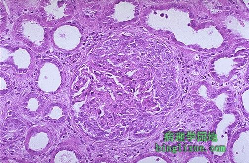
肾小球内由增生的壁层上皮细胞组成的新月体。因为展迅速,新月体性肾小球肾炎也称作快速进行性肾小球肾炎。它有多种病因,本例是由SLE引起。左下角的肾小球毛细血管管壁显著增厚(狼疮性肾炎时的“线圈”征)。 Seen here within the glomeruli are crescents composed of proliferating epithelial cells. Crescentic glomerulonephritis is known as rapidly progressive glomerulonephritis (RPGN) because this disease is very progressive. There are several causes, and in this case is due to SLE. Note in the lower left glomerulus that the capillary loops are markedly thickened (the so-called "wire loop" lesion of lupus nephritis). |
 |
壁层上皮细胞形成新月体性肾炎的肾小球,新月体从四周挤压毛细血管丛。快速进行性肾小球肾炎可以是特发的,也可以来自SLE、感染后肾小球肾炎(如在某些链球菌感染后的肾炎)或各种类型的血管炎及Goodpasture综合征(肺出血-肾炎综合征)。 Here is another glomerulus with epithelial crescents squashing the glomerular tufts from all sides. RPGN may be idiopathic or may result from SLE, post-infectious GN (as in some cases of post-streptococcal GN), from various types of vasculitis, and from Goodpasture's syndrome. |
 |
免疫荧光显示,肾小球抗纤维蛋白原抗体阳性。快速进展性肾小球肾炎时,肾小球损伤严重,以致纤维蛋白原都漏入了Bowman囊,导致上皮细胞的增生及新月体形成。 This immunofluorescence micrograph of a glomerulus demonstrates positivity with antibody to fibrinogen. With a rapidly progressive GN, the glomerular damage is so severe that fibrinogen leaks into Bowman's space, leading to proliferation of the epithelial cells and formation of a crescent |
 |
免疫荧光显微镜下,抗IgG抗体阳性。表现为光滑、弥漫分布的线性荧光。它是Goodpasture综合征的肾小球基底膜抗体的特点。 This immunofluorescence micrograph shows positivity with antibody to IgG has a smooth, diffuse, linear pattern that is characteristic for glomerular basement membrane antibody with Goodpasture's syndrome. |