
 |
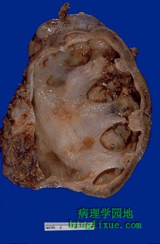
严重的肾盂积水,仅剩下一层薄薄的肾皮质。这种肾脏是没有功能的,它也是容易发生感染的地方。如果病变是单侧的,原因可能是从肾盂到输尿管口的病变。在本例,一个大的“鹿角”样(因外形象鹿角,故而名之)结石充满了肾盂和肾盏。如果病变是双侧的,原因可能是膀胱三角或尿道前列腺部的病变;或是其它能侵犯两侧输尿管的病变(如大的肿瘤块)。 Here is a kidney with much more advanced hydronephrosis in which there is only a thin rim of remaining renal cortex. Such a kidney is non-functional and a source for ongoing infection. If this process is unilateral, then the problem originates from the ureteral orifice up to the pelvis. In this case, a large "staghorn" calculus (so named because the prominent projections of the stone into the calyces resemble deer antlers) was present that filled up the pelvis and calyceal system. If this process were bilateral, then the problem would originate in the bladder trigone or urethra (or the prostate around the urethra) or some process (such as a large neoplasm) that could impinge on both ureters. |
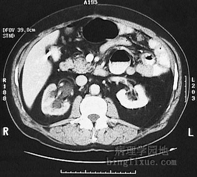 |
腹部增强CT显示输尿管阻塞导致右(图左侧)肾盂和输尿管积水。 This abdominal CT scan with contrast demonstrates right hydronephrosis and hydroureter as a consequence of ureteral obstruction. |
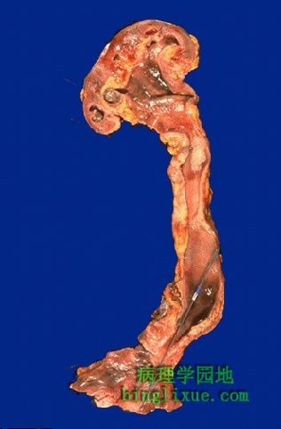 |
用金属探针指示了在输尿管口的长期阻塞(可能是先天性的)。此处明显肾盂积水和输尿管积水由阻塞引起。 A long-standing obstruction (probably congenital) at the ureteral orifice through which the metal probe passes led to the marked hydroureter and hydronephrosis seen here. |
 |
下腹部和骨盆部X线显示右尿路(图左侧),从右肾盂到膀胱。在右上象限(图左上)明亮的小块是先前胆囊手术留下的。 This radiograph of the lower abdomen and pelvis demonstrates a stent that has been placed in the urinary tract from the right renal pelvis to the bladder to relieve obstruction. (The bright radiopaque clips at the patient's upper right quadrant are left from previous cholecystectomy). |
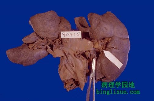 |
白色的箭头指示肾静脉血栓。肾静脉血栓形成有很多原因:创伤、受肿瘤压迫、肾细胞癌侵犯肾静脉、膜性肾小球肾炎引起的肾病综合征等。 The white arrow marks a renal vein thrombus. There are a host of etiologies for renal vein thrombosis, including trauma, compression by neoplasms, renal vein invasion by renal cell carcinoma, and nephrotic syndrome with membranous glomerulonephritis. |
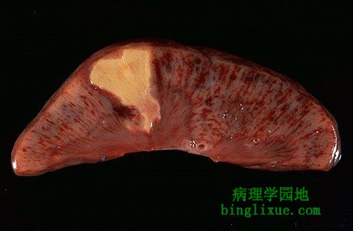 |
急性肾梗死,属凝固性坏死,坏死区域呈楔形,是由于组织血供减少引起缺血,终而产生灰白的梗死区。有少量从肾包膜来的血液供应紧邻的皮质。髓质和余下的皮质呈充血状。 This is an acute renal infarction. Note the wedge shape of this zone of coagulative necrosis resulting from loss of blood supply with resultant tissue ischemia that produces the pale infarct. The small amount of blood supply from the capsule supplies the immediate subcortical zone. The remaining cortex is congested, as is the medulla. |
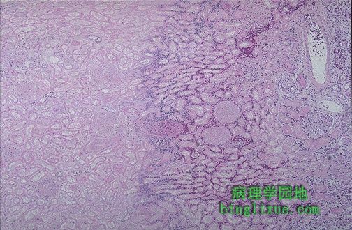 |
镜下显示急性肾梗死。最右边是正常肾脏,其左边是濒临坏死的充血区,再往左边是苍白、粉红的梗死区,此处肾小球和肾小管都已坏死。 This is the microscopic appearance of an acute renal infarct. At the far right is normal kidney, then to the left of that hyperemic kidney that is dying, then to the left of that pale pink infarcted kidney in which both tubules and glomeruli are dead. |
 |
尸检剖开膀胱显示充血粘膜,是急性膀胱炎的表现。 This bladder at autopsy has been opened to reveal areas of hyperemia of the mucosa. This is acute cystitis. |
 |
可见前列腺显著增大,不仅是两侧小叶,中叶也肥大。这导致了尿道前列腺部阻塞,引起慢性尿道梗阻。结果,每次排尿时为了克服阻力,膀胱代偿性肥大,膀胱表面形成小梁。另外,在膀胱内还有棕黄*色的结石。 The markedly enlarged prostate seen here has not only large lateral lobes, but a very large median lobe as well that obstructs the prostatic urethra and led to chronic urinary tract obstruction. As a result, the bladder became both enlarged and hypertrophied as it had to work against the obstruction with every episode of urination. That is why the surface of the bladder appears trabeculated. Note also that a yellowish-brown calculus formed in the bladder. |
 |
肾脏下极有一个1cm大小的灰黄*色脓肿。感染可以由尿路上行至肾(如由膀胱感染引起,称为逆行性感染,也叫上行性感染),也可以由脓毒血症时的血行播散(称为顺行性感染)而来。这种单一的脓肿可能是血行播散的。 In the lower pole of this kidney is a 1 cm pale yellow abscess. Infections can reach the kidney either by ascending up the urinary tract (from a bladder infection, for example) or by hematogenous spread with sepsis. This lone abscess was probably hematogenous in origin. |