
 |
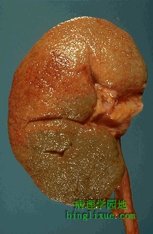
肾血管病导致的良性肾硬化,肾脏小动脉管壁增厚、管腔狭窄。高血压或糖尿病时常出现动脉玻璃样变。这导致局部肾实质减少形成片状缺血性萎缩,从而使得肾脏表面呈现特征性的颗粒状外观。 Here is an example of renal vascular disease known as benign nephrosclerosis. The smaller arteries in the kidney have become thickened and narrowed. Hyaline arteriolosclerosis with hypertension or diabetes mellitus is usually present. This leads to patchy ischemic atrophy with focal loss of parenchyma that gives the surface of the kidney the characteristic granular appearance as seen here. |
 |
恶性肾硬化时,肾脏出现局灶性出血。发生于恶性高血压(急进型高血压),血压很高(如:300/150mmHg)。 In malignant nephrosclerosis, the kidney demonstrates focal small hemorrhages. This is due to an accelerated phase of hypertension in which blood pressures are very high (such as 300/150 mm Hg). |
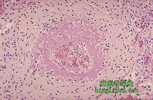 |
恶性高血压导致小动脉纤维素样坏死。动脉损伤引起粉红色纤维素坏死物形成,因此称为纤维素样坏死。 Malignant hypertension leads to fibrinoid necrosis of small arteries as shown here. The damage to the arteries leads to formation of pink fibrin--hence the term "fibrinoid". |
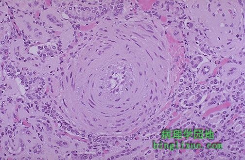 |
恶性高血压引起的动脉壁增厚发生增生性小动脉炎,小动脉呈洋葱皮样表现。 Thickening of the arterial wall with malignant hypertension also produces a hyperplastic arteriolitis. The arteriole has an "onion skin" appearance. |
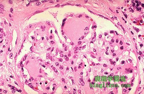 |
糖尿病性结节性肾小球硬化(kimmelstiel-wilson病)。肾小球毛细血管袢间有结节状粉红色玻璃样物。这是系膜间质显著增加的结果。系膜间质增加是蛋白非酶糖基化作用造成的。 This is nodular glomerulosclerosis (the Kimmelstiel-Wilson disease) of diabetes mellitus. Nodules of pink hyaline material form in regions of glomerular capillary loops in the glomerulus. This is due to a marked increase in mesangial matrix from damage as a result of non-enzymatic glycosylation of proteins |
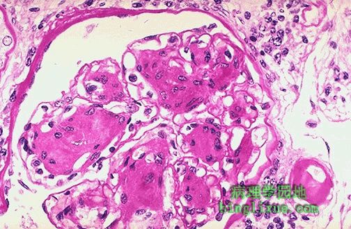 |
长期糖尿病病人结节性肾小球硬化的PAS染色。图右下的小动脉管壁显著增厚,是糖尿病肾病的玻璃样变性的小动脉的典型表现。 This is a PAS stain of nodular glomerulosclerosis (Kimmelstiel-Wilson disease) in a patient with long-standing diabetes mellitus. Note also the markedly thickened arteriole at the lower right which is typical for the hyaline arteriolosclerosis that is seen in diabetic kidneys as well. |
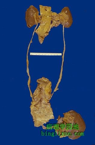 |
多种肾脏疾病,如:肾血管性疾病、肾小球肾炎或慢性肾盂肾炎等最终都可以发展为为终末期肾。如图显示终末期肾,双侧肾脏缩小,出现慢性肾衰,病人BUN(血尿素痰)和Cr(肌酐)升高。慢性肾衰进行透析治疗或肾移植。 The end result of many renal diseases--whether they are renal vascular diseases, glomerulonephritis, or chronic pyelonephritis--is end stage renal disease. In end stage renal disease, the kidneys are small bilaterally, as shown here. This condition is associated with chronic renal failure, and the patient's BUN and creatinine are elevated. Chronic renal failure can be treated by dialysis or by transplantation, as shown here. |
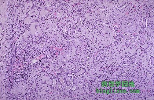 |
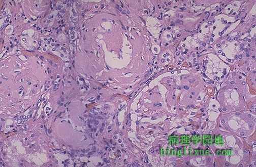
不管病因如何,终末期肾显微结构相似,因此肾活检在判断病因方面意义不大。它表现为皮质纤维化、肾小球硬化、慢性炎细胞弥散侵润、动脉壁增厚;部分肾小管常扩张并充满粉红色的胶样管型,形态与甲状腺滤泡相似。 The microscopic appearance of the "end stage kidney" is similar regardless of cause, which is why a biopsy in a patient with chronic renal failure yields little useful information. The cortex is fibrotic, the glomeruli are sclerotic, there are scattered chronic inflammatory cell infiltrates, and the arteries are thickened. Tubules are often dilated and filled with pink casts and give an appearance of "thyroidization." |
 |
肾脏淀粉样变性是一种能增加肾脏体积的慢性肾脏疾病。皮质有灰白色淀粉样沉积物,在图中上方最明显。 Here is a chronic renal disease that may actually increase the size of the kidney. This is amyloidosis. Pale deposits of amyloid are present in the cortex, most prominently at the upper center. |
 |
动脉壁及其周围、肾小球和肾间质可见无定形的粉红色淀粉样沉积物。刚果红染色淀粉样物呈粉红色。淀粉样物质沉积使肾脏体积增大,但功能下降。 The amorphous pink depositis of amyloid may be found in and around arteries, in interstitium, or in glomeruli. A Congo red stain will demonstrate the pink material to be amyloid. Such collections of amyloid add to renal bulk, but diminish renal function. |