
 |
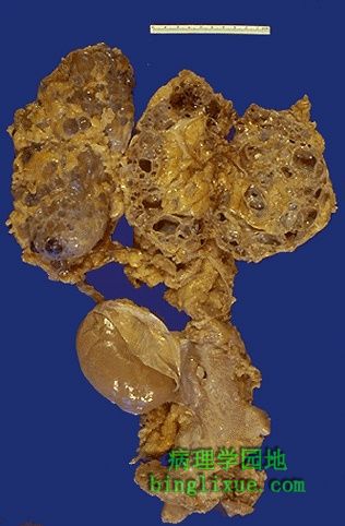
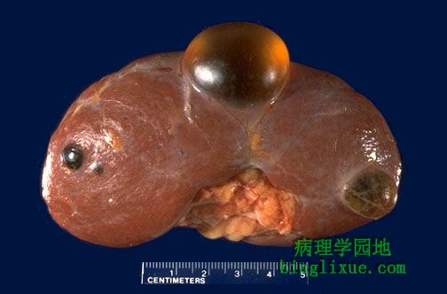
为了充分展示腹膜后双侧肿大的肾脏,尸解时去除了胸、腹腔脏器。这位显性遗传性多囊肾患者是成年人,死于慢性肾衰的并发症。 The contents of the chest and peritoneal cavity have been removed at autopsy here to reveal markedly bilaterally enlarged kidneys in the retroperitoneum in an adult who died from complications of chronic renal failure. This patient had dominant polycystic kidney disease (DPKD). |
 |
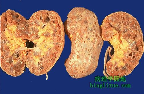
显性遗传性多囊肾患者的肾脏切面显示:实质为大囊肿取代,和正常的移植肾相比,有囊肿的肾脏多么大啊! The cut surfaces of these kidneys in a patient with DPKD reveal that the parenchyma is replaced by large cysts. Note how large these kidneys are in relation to the normal sized transplanted kidney. |
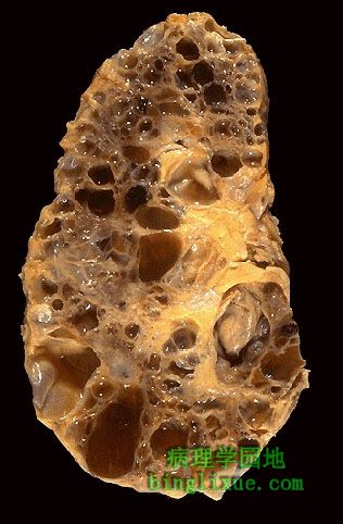 |
此患者肾脏竟然重达3公斤。本病呈显性遗传,所以家族发病率为50%。通常在出生时并没有囊肿,囊肿是在后来缓慢发展的。所以肾衰常在中、老年时发生。 This kidney in a patient with DPKD weighed 3 kilograms! This disease is inherited with an autosomal dominant pattern, so the recurrence risk in the family is 50%. The cysts are not usually present at birth, but develop slowly over time, so the onset of renal failure occurs in middle age to later adult life. |
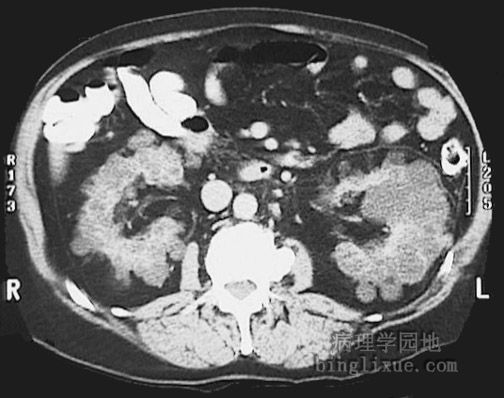 |
腹部横断面CT显示大的囊肿肾。 This CT scan of the abdomen demonstrates large cystic kidneys. |
 |
这些肾脏大小正常,有一些散在囊肿。但没有一个囊肿大于2cm。这是长期透析引起的囊肿。 These kidneys are about normal in size but have a few scattered cysts, none of which is over 2 cm in size. This is cystic change associated with chronic renal dialysis. |
 |
单纯性肾囊肿,可以是多发的,但它们从来没有多囊变化,它不会发展成慢性肾衰。 Simple renal cysts, as seen here, can also be multiple, but they are never as numerous as with polycystic change, and they do not predispose to chronic renal failure. |
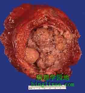 |
这是从一个有长期吸烟史的患者切下的膀胱。他曾有血尿。组织学证实膀胱内的肿块是移行细胞癌。尿道上皮的任一处都可能发生移行细胞癌,但膀胱多见。移行细胞癌是多灶性的,有复发倾向。 This bladder was removed surgically from a male who had a long history of smoking. He had presented with hematuria. The opened bladder reveals masses of a neoplasm that histologically proved to be urothelial carcinoma (TCC). TCC can arise anywhere in the urothelium, but is most common in bladder. TCC is often multifocal and has a tendency to recur. |
 |
被切除的肾脏切面展示了正常皮质和髓质,肾盂有局灶性的乳头状肿块,为移行细胞癌。 The cut surfaces of the kidney removed surgically here demonstrate normal cortex and medulla, but the calyces show focal papillary tumor masses of urothelial carcinoma. |
 |
这是另一例肾盂移行细胞癌。它侵袭性很强,侵入了肾实质。血尿是常见的表现。 Here is another example of urothelial carcinoma that is more aggressive and is invading into the renal parenchyma. Hematuria is a frequent presenting symptom. |
 |
低倍镜下显示移行细胞癌。肿瘤表面的叶子样乳头状突起突向左侧。分化较好,类似于尿道上皮,不过确实是肿瘤,未见向右侧浸润。 A urothelial carcinoma of the urothelium is shown here at low power to reveal the frond-like papillary projections of the tumor above the surface to the left. It is differentiated enough to resemble urothelium, but is a mass. No invasion to the right is seen at this point. |