
 |
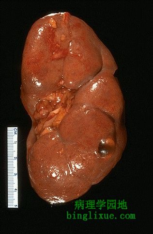
正常成年人肾脏,已去除肾包膜。肾脏仍保持着胚胎时的小叶形态(成年人有时保留着胚胎时的小叶形态)。位于左侧中部的肾门处有一些脂肪组织。右下有一个表面光滑、充满清亮液体的单纯性肾脏小囊肿。囊肿可以单发,也可散在于肾脏实质周围。囊肿在成年人并不罕见。 Here is a normal adult kidney. The capsule has been removed and a pattern of fetal lobulations still persists, as it sometimes does. The hilum at the mid left contains some adipose tissue. At the lower right is a smooth-surfaced, small, clear fluid-filled simple renal cyst. Such cysts occur either singly or scattered around the renal parenchyma and are not uncommon in adults. |
||||||||||||
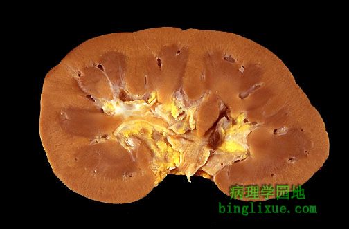 |
横切面显示成年人肾脏外周皮质颜色较浅,髓质及中间的肾盂色较暗。 In cross section, this normal adult kidney demonstrates the lighter outer cortex and darker medulla with central pelvis. |
||||||||||||
 |
静脉肾盂造影显示正常泌尿道,可见肾盂、输尿管和膀胱。 This intravenous pyelogram (IVP) of a normal urinary tract on the left demonstrates contrast filling the pelvis, ureter, and bladder. |
||||||||||||
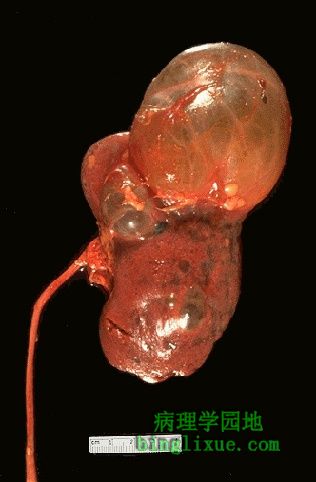 |
肾脏上极更大的单纯性肾囊肿,还有一些小囊肿散布于肾脏。左侧输尿管通向南面。放射影像学检查可以发现这样大小的囊肿。根据其囊内液体的密度可以把它和肿瘤区别开来。 Here is a much larger simple renal cyst of the upper pole. Other smaller cysts are also scattered around the kidney. The ureter exits south on the left. Such a large renal cyst would be seen on a radiographic imaging procedure, but could probably be distinguished from a neoplasm by its fluid density. |
||||||||||||
 |
腹部横断面CT显示右肾(图左)单纯性大囊肿,左肾(图右)可见小囊肿。 This CT scan of the abdomen demonstrates a large simple renal cyst on the right and a smaller cyst on the left. |
||||||||||||
 |
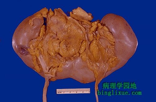
可见双侧肾脏都有两个输尿管,通向膀胱(已剖开)。表面光滑的正常两肾间,可见一小段主动脉。1/150的人会出现部分或全部的单侧或双侧的双输尿管。因为尿流的异常及两个输尿管在膀胱的开口接近,可能导致阻塞。但总的来说,双输尿管是偶然发现的(泌尿科医生除外)。 Double ureters are seen exiting from each kidney and extending to the bladder that has been opened. A small segment of aorta is seen between the normal, smooth-surfaced kidneys. A partial or complete duplication of one or both ureters occurs in about 1 in 150 persons. There is a potential for obstructive problems due to the abnormal flow of urine and the entrance of two ureters into the bladder in close proximity, but most of the time this is an incidental finding (except to a urologist). |
||||||||||||
 |
马蹄肾,是一种先天性的异常,常和其它异常或特殊基因缺陷而引起的综合征有关。也可单独发生。可能是输尿管穿过肾脏组织“桥”时的路径异常造成的。这样可能导致部分梗阻而致肾盂积水。 Here is a "horseshoe" kidney. This is a congenital anomaly that most often occurs in association with other anomalies or syndromes with specific genetic defects such as trisomy 18. However, it can also occur as an isolated anomaly. The possible problem here is that the ureters take an abnormal course across the "bridge" of renal tissue and this can lead to partial obstruction with hydronephrosis. |
||||||||||||
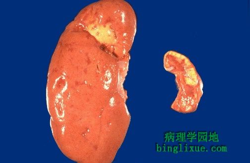 |
左侧为相对正常的右肾,仅有少量散在的浅的皮质瘢痕,肾上极有大的黄褐色瘢痕。左肾(右侧)因肾动脉梗阻而萎缩了,可导致高血压(Goldblatt 肾)。 There is a relatively normal kidney at the left with only a few scattered, shallow cortical scars and one fairly large pale tan-yellow scar in the upper pole. The left kidney is atrophic because of renal arterial occlusion. Such a situation can lead to hypertension (Goldblatt kidney). |
||||||||||||
 Stones containing calcium are far more frequent than other types, and about half the time occur when there is hypercalciuria. Only about 10% of the time do they appear as a consequence of hypercalcemia. The struvite stones are also known as "infection" stones because bacteria such as Proteus that split urea to ammonia favor their formation. Uric acid stones may be seen in association with gout, but often are not, and may just reflect increased precipitation of urates in an acid urine. Rare cystine stones also form in acid urine. |
结石经过输尿管的模式图。当钙等溶质排出增多,尿的酸、碱度及郁积情况或溶质浓度适合时,就会形成结石。形成结石的物质多种多样,其中磷酸钙、草酸钙结石较多。尿路结石通常是单侧的,直径多为1-3mm,导致疼痛、血尿,当然大结石可导致肾盂积水或输尿管积水。 The passage of a calculus (stone) through the urinary tract is diagrammed here. Calculi form when there is increased excretion of solutes such as calcium and when urine alkalinity, acidity, stasis, and/or concentration are favorable. The most common varieties of calculi are:
|
||||||||||||
 |
大的肾结石阻塞了肾脏下极的肾盏,导致了局部的肾盂积水。梗阻、扩张引起的肾盂积水导致了感染。扩张肾盏的表面粘膜充血,靠近扩张肾盏的区域为黄褐苍白区,这是炎症感染的特征。肾上极正常,皮、髓质分界清楚。 There was a large renal calculus (stone) that obstructed the calyces of the lower pole of this kidney, leading to a focal hydronephrosis (dilation of the collecting system). The stasis from the obstruction and dilation led to infection. The infection with inflammation is characterized by the pale yellowish-tan areas next to the dilated calyces with hyperemic mucosal surfaces. The upper pole is normal and shows good corticomedullary demarcations. |