
 |
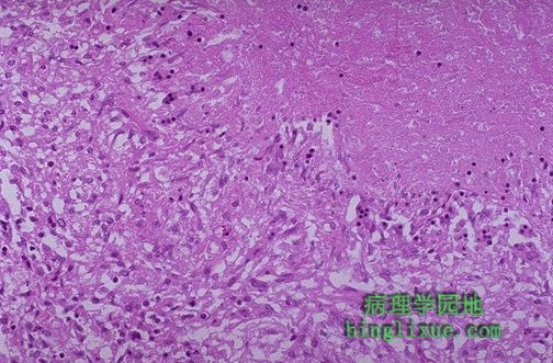
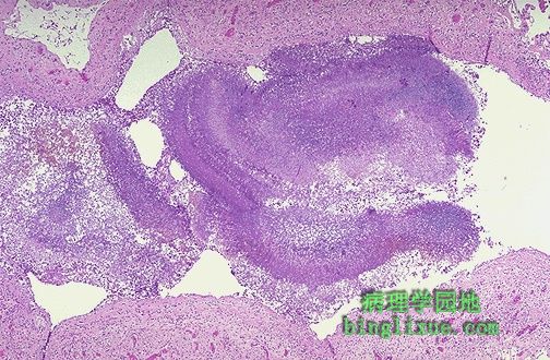
高倍镜示肉芽肿边缘。右上部是无定形的粉红色干酪样坏死物,它由肉芽肿坏死成分以及感染的微生物构成。此区由炎症成分围成环状,这些成分包括上皮样细胞,淋巴细胞,成纤维细胞。 The edge of a granuloma is shown here at high magnification. At the upper right is amorphous pink caseous material composed of the necrotic elements of the granuloma as well as the infectious organisms. This area is ringed by the inflammatory component with epithelioid cells, lymphocytes, and fibroblasts. |
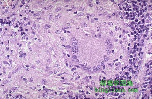 |
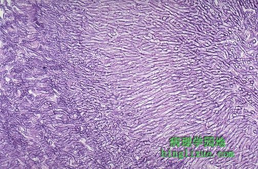
高倍镜下,肉芽肿内可见上皮样细胞,胞核染色较浅、胞质粉红色。此种细胞融合成团,称巨细胞。感染性肉芽肿形成典型的巨细胞叫朗格罕斯巨细胞,它的细胞核沿细胞边缘排列。肉芽肿性炎持续数月到数年。 At high magnification, the granuloma demonstrates that the epithelioid macrophages are elongated with long, pale nuclei and pink cytoplasm. The macrophages organize into committees called giant cells. The typical giant cell for infectious granulomas is called a Langhans giant cell and has the nuclei lined up along one edge of the cell. The process of granulomatous inflammation takes place over months to years (did you ever hear of a committee action that was completed in a short time?) |
 |
为了在组织切片上发现分支杆菌,进行抗酸染色( AFB 染色)。高倍镜示,分支杆菌被染成红棒。 In order to find the mycobacteria in a tissue section, a stain for acid fast bacilli is done (AFB stain). The mycobacteria stain as red rods, as seen here at high magnification. |
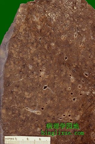 |
免疫反应低下或由广泛感染受抑时,就可能看到图示的肉芽肿表现。因为肺实质内存在大量散在的小棕褐色肉芽肿,直径约 2~4 mm ,称为粟粒性肉芽肿。 When the immune response is poor or is overwhelmed by an extensive infection, then it is possible to see the gross pattern of granulomatous disease seen here. This is a "miliary" pattern of granulomas because there are a multitude of small tan granulomas, about 2 to 4 mm in size, scattered throughout the lung parenchyma. The miliary pattern gets its name from the resemblence of the granulomas to millet seeds. |
 |
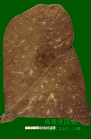
近距离见分布于全肺的粟粒性肉芽肿。感染性播散在其它器官中也可以产生类似的病变。 At closer range, the miliary pattern is seen throughout the lung. Dissemination of the infectious agent (M. tuberculosis, fungi) may produce a similar pattern in other organs. |
 |
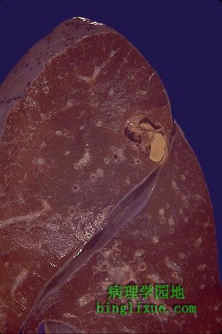
曲霉菌引起的真菌性肉芽肿。损害轻易穿过裂隙暗示存在传染。肿块最初通常被组织膜阻碍。这个肉芽肿不规则,边缘为红色,以及中心质地坚韧,桔黄*色。 This is a fungal granuloma produced by Aspergillus. An infectious process is suggested by the fact that the lesion has crossed the fissure as though it weren't there. A neoplasm usually is initially impeded by an anatomical barrier. This granuloma has an irregular, red margin and a firm, tan-orange center. |
 |
另例真菌性肉芽肿。 Here is another fungal granuloma. |
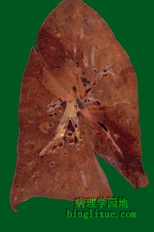 |
肺受真菌性肉芽肿影响较广。真菌感染常见于免疫抑制。 The lung involvement by these fungal granulomas is more extensive. Fungal infections are more common in patients who are immunosuppressed. |
 |
支气管处可见由蓝色的菌丝构成的真菌球。当真菌在因结核损害形成的腔洞内植入时,就可能形成真菌球。 A fungus ball composed of blue-staining hyphal elements of Aspergillus is seen here in a bronchus. Fungus balls may also form when fungi colonize cavitary lesions of tuberculosis. |
 |
曲霉肿中有分支、有隔膜的菌丝聚集并呈放射状排列。 Branching, septate hyphae are close-packed here and radiating outward in this aspergilloma. |