
 |
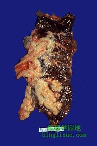
中央型小细胞(燕麦细胞)癌向周围蔓延。肿瘤切面质地软,呈片状,灰白色。图示肿瘤导致左主支气管阻塞,因此远端肺组织塌陷。燕麦细胞癌具有很强的侵袭性,原发瘤不是很大就已经发生了广泛转移。 Arising centrally in this lung and spreading extensively is a small cell anaplastic (oat cell) carcinoma. The cut surface of this tumor has a soft, lobulated, white to tan appearance. The tumor seen here has caused obstruction of the main bronchus to left lung so that the distal lung is collapsed. Oat cell carcinomas are very aggressive and often metastasize widely before the primary tumor mass in the lung reaches a large size. |
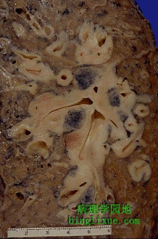 |
正沿支气管蔓延的燕麦细胞癌。斑点状黑色圆形区域说明存在肺门淋巴结转移。对化疗敏感,比放射治疗或外科手术治疗的疗效要好,但是预后较差。燕麦细胞癌几乎只发生于吸烟者。 Here is an oat cell carcinoma which is spreading along the bronchi. The speckled black rounded areas represent hilar lymph nodes with metastatic carcinoma. These neoplasms are more amenable to chemotherapy than radiation therapy or surgery, but the prognosis is still poor. Oat cell carcinomas occur almost exclusively in smokers. |
 |
小细胞(燕麦细胞)癌细胞呈暗蓝色,挤压后聚集成片状,细胞质少。 This is the microscopic pattern of a small cell anaplastic (oat cell) carcinoma in which small dark blue cells with minimal cytoplasm are packed together in sheets. |
 |
两例错构瘤,为肺良性肿块。胸片上这些不常见的损害(硬币损害)可作为与肉芽肿及局限化的恶性肿瘤鉴别诊断的依据。质地坚韧,X线片上可见钙化区。绝大多数比较小(小于 2 厘米)。 Here are two examples of a benign lung neoplasm known as a pulmonary hamartoma. These uncommon lesions appear on chest radiograph as a "coin lesion" that has a differential diagnosis of granuloma and localized malignant neoplasm. They are firm and discreet and often have calcifications in them that also appear on radiography. Most are small (less than 2 cm). |
 |
显微镜下,肺错构瘤主要由良性软骨(右侧)构成,它与间质混在一起,左边是散在的支气管腺体。错构瘤是发生于器官的肿块,由所在部位的正常组织成分构成,但形成了不规则肿块。 The pulmonary hamartoma is seen microscopically to be composed mostly of benign cartilage on the right that is jumbled with a fibrovascular stroma and scattered bronchial glands on the left. A hamartoma is a neoplasm in an organ that is composed of tissue elements normally found at that site, but growing in a haphazard mass. |
 |
可见肺表面散大的多个大小不一的肿块。灰白色结节状肿块是转移癌的特征。肺转移瘤比原发性肺肿瘤更常见,因为许多其它原发肿瘤可通过血道转移到肺。甚至图示的肺门的结节也是转移癌的结节。瘤结节通常出现在边缘,阻塞作用小。 Multiple variably-sized masses are seen in all lung fields. These tan-white nodules are characteristic for metastatic carcinoma. Metastases to the lungs are more common even than primary lung neoplasms simply because so many other primary tumors can metastasize to the lungs. Even the hilar nodes in this photograph demonstrate nodules of metastatic carcinoma. The nodules are usually in the periphery and do not cause major obstruction. |
 |
胸部X线显示结肠腺癌转移到肺,肺出现多个转移性小结节。 This chest radiograph demonstrates a nodular pattern resulting from multiple small metastases to the lung from a colonic adenocarcinoma. |
 |
肺内体积较大但大小不一的癌转移性结节。 Here are larger but still variably-sized nodules of metastatic carcinoma in lung. |
 |
结肠腺癌转移到肺晚期的胸部X线表现。该病人同上面的是一个病人,只不过处于晚期。颈部见到的阴影是癌转移引起的颈椎病理性骨折手术修复时拧入的钢板。 This chest radiograph demonstrates a nodular pattern resulting from multiple metastases to the lung from a colonic adenocarcinoma. This is the same patient as the previous radiograph, but at a later point in the course. (The plate and screws in the cervical spine repaired a pathologic fracture from metastasis). |
 |
肺扩张的淋巴管可见来自乳腺浸润性导管癌转移灶。癌经常通过淋巴道转移。前列腺腺癌通过淋巴道转移到肺,肺叶之间和胸膜下的淋巴管中都可见癌细胞。 A nest of metastatic infiltrating ductal carcinoma from breast is seen in a dilated lymphatic channel in the lung. Carcinomas often metastasize via lymphatics. Prostatic adenocarcinoma is famous for metastasizing to the lungs in a "lymphangitic" pattern in which streaks of tumor appear between lung lobules and beneath the pleura in lymphatic spaces. |