
 |
尸体解剖显示胸腔和腹腔。胸腔内的肺为正常的粉红色饱满状,肺内只有极少的炭末沉着,因为这个80岁的男性从来不吸烟,并且不允许其它人在他的工作地点吸烟。纵隔内主要是脂肪。心包未被打开。横膈穹窿向上延伸到第6 肋水平。 The chest and abdominal cavities are opened here at autopsy. The lungs in the chest have a normal pink aerated appearance with minimal anthracotic pigmentation, because this 80 year old male never smoked and never allowed smoking in his workplace. The mediastinum contains mostly fat. The pericardial sac around the heart has not been opened.The diaphragmatic domes extend upward to the level of the 6th ribs. |
 |
正常肺的横切面(仅在右下肺的背面出现很少充血)。肺门淋巴结比较小,内部有大量炭末(吸入的炭末被肺巨噬细胞清除,并通过淋巴管,在淋巴结聚集。),使其呈现灰黑色。 This is a cross-section of normal lung (with only minimal posterior congestion at the lower right). The hilar lymph nodes are small and have enough anthracotic pigment (from dusts in the air breathed in, scavenged by pulmonary macrophages, transferred to lymphatics, and collected in lymph nodes) to make them appear greyish-black. |
 |
光滑、有光泽的脏层胸膜表面。严重肺水肿病人,肺小叶间淋巴液增多,因此肺小叶轮廓呈白色。 The smooth, glistening pleural surface of a lung is shown here. This patient had marked pulmonary edema, which increased the fluid in the lymphatics that run between lung lobules. Thus, the lung lobules are outlined in white. |
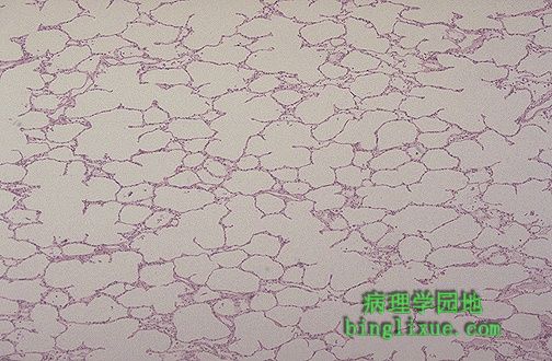 |
正常肺显微镜显示肺泡壁比较薄弱。肺泡内充满空气,有肺巨噬细胞(II型上皮细胞)。 This is normal lung microscopically. The alveolar walls are thin and delicate. The alveoli are well-aerated and contain only an occasional pulmonary macrophage (type II pneumonocyte). |
 |
高倍镜显示,肺泡内充满表面光滑并且泡沫状粉红色物质,这是肺水肿的特征。同时也要注意到肺泡壁毛细血管充血,血管内有许多红细胞。肺淤血和肺水肿在心衰病人身上和肺炎区域比较常见。 At high magnification, the alveoli in this lung are filled with a smooth to slightly floccular pink material characteristic for pulmonary edema. Note also that the capillaries in the alveolar walls are congested with many red blood cells. Congestion and edema of the lungs is common in patients with heart failure and in areas of inflammation of the lung. |
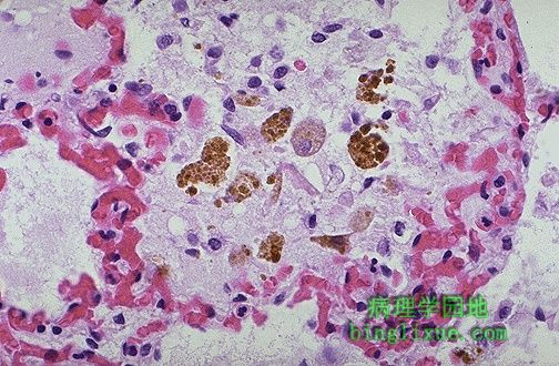 |
伴有毛细血管扩张的肺充血和血液流入肺泡腔,共同引起内部充满含铁血黄素的巨噬细胞增多,如图所示,红细胞破裂形成的棕色含铁血黄素颗粒出现在巨噬细胞的胞质内。由于与充血性心力衰竭有关,因此也称为心力衰竭细胞。 Pulmonary congestion with dilated capillaries and leakage of blood into alveolar spaces leads to an increase in hemosiderin-laden macrophages, as seen here. Brown granules of hemosiderin from break down of RBC's appear in the macrophage cytoplasm. These macrophages are sometimes called "heart failure cells" because of their association with pulmonary congestion with congestive heart failure. |
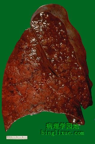 |
肺切面显示典型的灰黄*色实变区域的支气管肺炎。由于明显的肺充血,残余的肺组织呈暗红色。支气管肺炎(小叶性肺炎)出现典型的不规则实变病灶,病变以肺小叶为中心。图片左下部左下肺叶显示多病灶融合。实变区域比周围的肺组织坚韧。 The cut surface of this lung demonstrates the typical appearance of a bronchopneumonia with areas of tan-yellow consolidation. Remaining lung is dark red because of marked pulmonary congestion. Bronchopneumonia (lobular pneumonia) is characterized by patchy areas of pulmonary consolidation. These areas become almost confluent in the left lower lobe on the bottom left of the photograph.The areas of consolidation are firmer than the surrounding lung. |
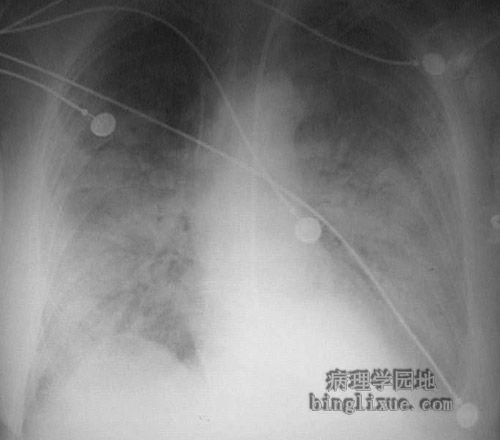 |
X线显示的不规则病灶与细菌性支气管肺炎病变相一致。典型的病原微生物有:肺炎链球菌、金黄*色葡萄球菌、绿浓杆菌、流感嗜血杆菌、肺炎克雷白杆菌等。 This radiograph demonstrates patchy infiltrates consistent with a bronchopneumonia from a bacterial infection. Typical organisms include Streptococcus pneumoniae, Staphylococcus aureus, Pseudomonas aeruginosa, Hemophilus influenzae, Klebsiella pneumoniae, among others. |
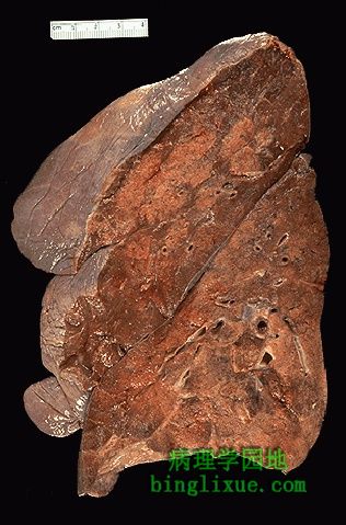 |
另例支气管肺炎。切面上看似比周围肺组织高一些的浅色区域,是肺实变区。 Here is another example of a bronchopneumonia. The lighter areas that appear to be raised on cut surface from the surrounding lung are the areas of consolidation of the lung. |
 |
放大后可见支气管肺炎病灶的不规则分布。实变区域范围很接近肺小叶(因此又称小叶性肺炎)。支气管肺炎是很典型的“医院感染型”肺炎,发生于已患其它疾病的病人。典型的病原微生物包括:金黄*色葡萄球菌, 克雷白菌属,大肠杆菌(E. coli),假单胞菌。 At higher magnification, the pattern of patchy distribution of a bronchopneumonia is seen. The consolidated areas here very closely match the pattern of lung lobules (hence the term "lobular" pneumonia).A bronchopneumonia is classically a "hospital acquired" pneumonia seen in persons already ill from another disease process. Typical bacterial organisms include: Staphylococcus aureus, Klebsiella, E. coli, Pseudomonas. |