
 |
食管鳞状细胞癌病人常有吸烟史和(或)酒精中毒史,然而食管腺癌常有Barrett食管。图示胃食管交界处有性质不明的肿块,可见溃疡形成和不规则黏膜,临床表现为疼痛和吞咽困难。 A history of smoking and/or alcoholism is often present in patients with esophageal squamous carcinoma, while a history of Barrett's esophagus precedes development of esophageal adenocarcinoma in many cases. Here, an ill-defined mass at the gastroesophageal junction produces mucosal ulceration and irregularity, which led to the clinical symptoms of pain and difficulty swallowing. |
 |
不规则微红色外生性溃疡,位于食管中段粘膜表面的的肿块是鳞状细胞癌。食管鳞状细胞癌的危险因素在美国包括大量吸烟和酒精中毒。在别的国家饮食因素可能起作用。 This irregular reddish, ulcerated exophytic mid-esophageal mass as seen on the mucosal surface is a squamous cell carcinoma. Risk factors for esophageal squamous carcinoma include mainly smoking and alcoholism in the U.S. In other parts of the world dietary factors may play a role. |
 |
胃镜可见伴溃疡的食管中段鳞状细胞癌引起食管腔狭窄。 Endoscopic views of an ulcerated mid-esophageal squamous cell carcinoma causing lumenal stenosis are seen. |
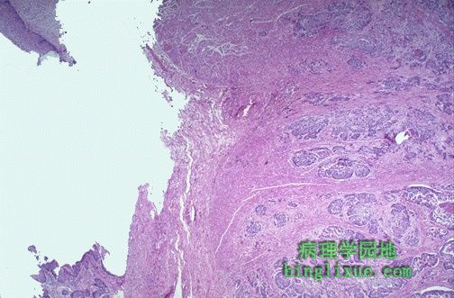 |
左上方是残留的食管鳞状上皮,已被食管中段鳞状细胞癌破坏。左侧可见肿瘤细胞巢正向下浸润突破黏膜下层。食管癌经常浸润破坏周围组织,使得外科手术切除很困难。 At the upper left is a remnant of squamous esophageal mucosa that has been undermined by an infiltrating squamous cell carcinoma of the mid-esophagus. Solid nests of neoplastic cells are infiltrating down through the submucosa at the right. Esophageal cancers often spread to surrounding structures, making surgical removal difficult. |
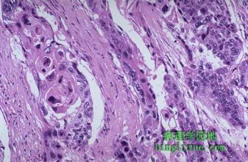 |
高倍镜下具有侵袭性的细胞巢中有丰富的粉红色细胞质,并且细胞边界清楚,呈现出鳞状细胞癌的典型特征。食管癌早期不易发现,因此预后不良。 At high power, these infiltrating nests of neoplastic cells have abundant pink cytoplasm and distinct cell borders typical for squamous cell carcinoma. Esophageal carcinomas are not usually detected early and, therefore, have a very poor prognosis. |
 |
正常胃外观,已沿着胃大弯剪开。食管在左边。在底部可见胃小弯。胃窦紧今幽门,幽门通向右下方的十二指肠。 This is the normal appearance of the stomach, which has been opened along the greater curvature. The esophagus is at the left. In the fundus can be seen the lesser curvatur. Just beyond the antrum is the pylorus emptying into the first portion of duodenum is at the lower right. |
 |
正常的胃底部的外观在上消化道内窥镜检查显示如图左侧。右侧是正常的十二指肠。 The normal appearance of the gastric fundus on upper GI endoscopy is shown at the left, with the normal duodenal appearance at the right. |
 |
中央偏右是正常的胃窦延伸至幽门的外观。十二指肠的起始段(十二指肠球部)在右边较远处。 This is the normal appearance of the gastric antrum extending to the pylorus at the right of center. The first portion of the duodenum (duodenal bulb) is at the far right. |
 |
内窥镜图里可见正常的幽门在左边,十二指肠的起始部在右边。 In the endoscopic views, the normal appearance of the pylorus is seen at the left, with the first portion of the duodenum at the right. |
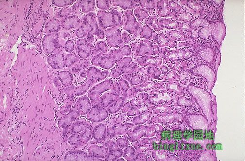 |
胃底正常黏膜,小凹由苍白的柱状黏液细胞排列形成长的腺体,此腺体包含能够分泌盐酸的、亮粉红色的壁细胞。 This is the normal appearance of the gastric fundal mucosa, with short pits lined by pale columnar mucus cells leading into long glands which contain bright pink parietal cells that secrete hydrochloric acid. |