
 |
直肠镜检。 Rectum |
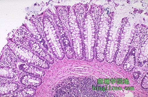 |
正常结肠黏膜。表明腺管由很多杯状细胞排列。黏膜下层有淋巴结。内脏相关的淋巴组织作为一个整体代表着体内最大的淋巴器官。 This is normal colonic mucosa. Note the crypts that are lined by numerous goblet cells. In the submucosa is a lymphoid nodule. The gut-associated lymphoid tissue as a unit represents the largest lymphoid organ of the body. |
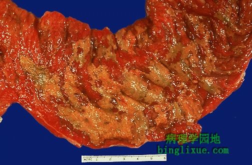 |
假膜性肠炎,图示结肠黏膜表面充血,部分由黄绿色渗出物覆盖。黏膜本身并没有被侵蚀。广谱抗生素的应用(例如氯林可霉素)和/或免疫抑制剂都使得细菌例如梭状芽孢杆菌属或S. Aureus,或真菌类例如念球菌属的过度生长,引起这种表现。 This is an example of pseudomembranous enterocolitis. The mucosal surface of the colon seen here is hyperemic and is partially covered by a yellow-green exudate. The mucosa itself is not eroded. Broad spectrum antibiotic usage (such as clindamycin) and/or immunosuppression allows overgrowth of bacteria such as Clostridium dificile or S. aureus or fungi such as Candida to cause this appearance. |
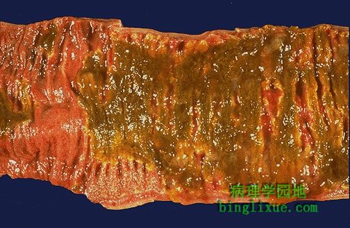 |
回肠假膜性炎。黄绿色的渗出物覆盖了大部分的黏膜。 This is another example of pseudomembranous inflammation, this time in the ileum. A greenish-yellow exudate covers most of the mucosal surface. |
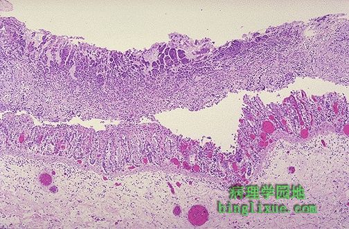 |
显微镜下,假膜由炎细胞、坏死的上皮细胞和有微生物过度生长的黏液组成,黏膜下显示的是充血而完整的血管。 Microscopically, the pseudomembrane is seen to be composed of inflammatory cells, necrotic epithelium, and mucus in which the overgrowth of microorganisms takes place. The underlying mucosa shows congested vessels, but is still intact. |
 |
左边覆盖的假膜有很多的炎症细胞,主要是中性粒细胞。 At higher magnification, the overlying pseudomembrane at the left has numerous inflammatory cells, mainly neutrophils. |
 |
正常阑尾,背景为盲肠。 This is the normal appearance of the appendix against the background of the cecum. |
 |
在下面可见阑尾口在盲肠的两个叉或袋状的折叠之间的结肠镜检图。 The colonoscopic view of the appendiceal orifice between the fork of two haustral folds in the cecum is seen here. |
 |
外科切除阑尾,病人最初主要是腹痛,但后来主要定位在右下腹,体格检查显示右下腹反跳痛阳性4+。白细胞计数提高到11500。图示急性阑尾炎有黄褐色渗出物和充血,在阑尾周围有过量的脂肪,而不是光滑的、发亮的浅褐色浆膜表面。 This appendix was removed surgically. The patient presented with abdominal pain that initially was generalized, but then localized to the right lower quadrant, and physical examination disclosed 4+ rebound tenderness in the right lower quadrant. The WBC count was elevated at 11,500. Seen here is acute appendicitis with yellow to tan exudate and hyperemia, including the periappendiceal fat superiorly, rather than a smooth, glistening pale tan serosal surface. |
 |
急性阑尾炎病人的阑尾远端。阑尾已经被纵切成两半。左侧显示浆膜表面,有黄褐色渗出物。右侧显示切面,有黄褐色的粘膜渗出物并有有边缘充血。 This is the tip of the appendix from a patient with acute appendicitis. The appendix has been sectioned in half. The serosal surface at the left shows a tan-yellow exudate. The cut surface at the right demonstrates yellowish-tan mucosal exudation with a hyperemic border. |