
 |
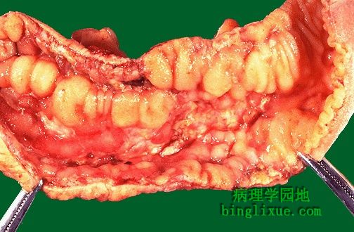
Crohn病发生在小肠。黏膜表面有不规则的充血结节和浅表溃疡灶。 This is another example of Crohn's disease involving the small intestine. Here, the mucosal surface demonstrates an irregular nodular appearance with hyperemia and focal superficial ulceration. |
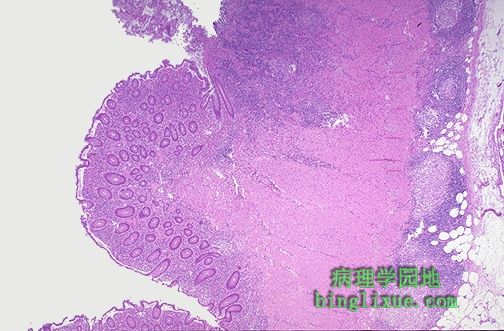 |
显微镜下Crohn病的特点是穿壁性炎症。炎细胞(兰色浸润)从黏膜层到黏膜下层、肌层均有浸润,在浆膜表面苍白的肉芽肿中心出现结节性浸润。 Microscopically, Crohn's disease is characterized by transmural inflammation. Here, inflammatory cells (the bluish infiltrates) extend from mucosa through submucosa and muscularis and appear as nodular infiltrates on the serosal surface with pale granulomatous centers. |
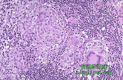 |
Crohn病炎性肉芽肿,有上皮样细胞、巨细胞和许多淋巴细胞。微生物特殊染色阴性。 At high magnification the granulomatous nature of the inflammation of Crohn's disease is demonstrated here with epithelioid cells, giant cells, and many lymphocytes. Special stains for organisms are negative. |
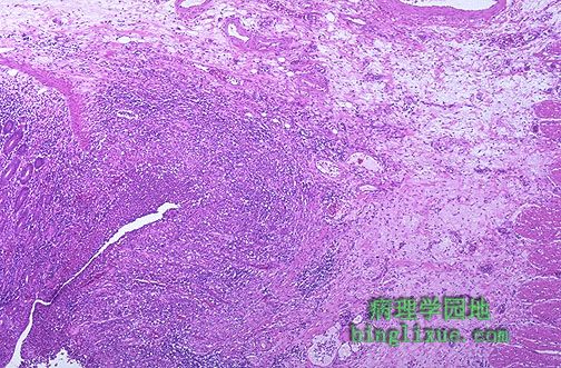 |
Crohn病并发症瘘管。左边可见裂缝从黏膜延伸到黏膜下层、肌层,最终形成瘘管。瘘管可在肠袢间、膀胱和皮肤形成。累及结肠的直肠周瘘管常见。 One complication of Crohn's disease is fistula formation. Seen here is a fissure extending through mucosa at the left into the submucosa toward the muscular wall, which eventually will form a fistula. Fistulae can form between loops of bowel, bladder, and skin. With colonic involvement, perirectal fistulae are common. |
 |
溃疡性结肠炎,严重的炎症从右下方的乙状结肠沿升结肠向上蔓延。在左下方是回肠末段的回盲瓣,不包括回肠末段。溃疡性结肠炎趋向于持续沿黏膜表面浸润和趋向于在直肠开始。黏膜遭受破坏,如图显示的仅仅是残留的岛屿状黏膜,称假息肉。 This gross appearance is characteristic for ulcerative colitis. The most intense inflammation begins at the lower right in the sigmoid colon and extends upward and around to the ascending colon. At the lower left is the ileocecal valve with a portion of terminal ileum that is not involved. Inflammation with ulcerative colitis tends to be continuous along the mucosal surface and tends to begin in the rectum. The mucosa becomes eroded, as in this photograph, which shows only remaining islands of mucosa called "pseudopolyps". |
 |
假息肉清晰可见发红炎性的岛屿状粘膜。假息肉间仅存肌层。 At higher magnification, the pseudopolyps can be seen clearly as raised red islands of inflamed mucosa. Between the pseudopolyps is only remaining muscularis. |
 |
广泛的溃疡性结肠炎,左边较低处可见回盲瓣。瓣膜上方开始带有红斑和颗粒性的粘膜炎症。随着病程的发展,粘膜损害发生融合,破坏残留的粘膜以至形成线性溃疡。 Here is another example of extensive ulcerative colitis (UC). The ileocecal valve is seen at the lower left. Just above this valve in the cecum is the beginning of the mucosal inflammation with erythema and granularity. As the disease progresses, the mucosal erosions coalesce to linear ulcers that undermine remaining mucosa. |
 |
可见到并不严重的溃疡性结肠炎的结肠镜检图,易碎的红斑粘膜,伴结肠带减少。 Colonoscopic views of less severe UC are seen below, with friable, erythematous mucosa with reduced haustral folds. |
 |
严重的溃疡性结肠炎病历中可见假息肉。残留的黏膜已经溃烂脱落,充血。 Pseudopolyps are seen here in a case of severe ulcerative colitis. The remaining mucosa has been ulcerated away and is hyperemic. |
 |
急性溃疡性结肠炎的结肠镜检图,但并不是被侵蚀得很厉害以至产生假息肉。 A colonoscopic view of active ulcerative colitis, but not so eroded as to produce pseudopolyps, is seen here. |