
 |
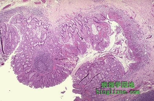
显微镜,溃疡性结肠炎最初限制在黏膜,溃疡破坏局部粘膜同时逐渐破坏周围粘膜。 Microscopically, the inflammation of ulcerative colitis is confined primarily to the mucosa. Here, the mucosa is eroded by an ulcer that undermines surrounding mucosa. |
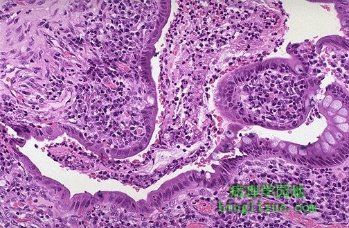 |
高倍镜下见严重的黏膜炎症。结肠黏膜上皮表明缺少杯状细胞,表面上有渗出物。急慢性炎症细胞都存在。 At higher magnification, the intense inflammation of the mucosa is seen. The colonic mucosal epithelium demonstrates loss of goblet cells. An exudate is present over the surface. Both acute and chronic inflammatory cells are present. |
 |
急性溃疡性结肠炎结肠显示粘膜隐窝有小脓肿,腺腔内可见中性粒细胞渗出 。黏膜下层有严重的炎症。不典型炎症中腺体缺少杯状细胞和细胞核深染。 The colonic mucosa of active ulcerative colitis shows "crypt abscesses" in which a neutrophilic exudate is found in glandular lumens. The submucosa shows intense inflammation. The glands demonstrate loss of goblet cells and hyperchromatic nuclei with inflammatory atypia. |
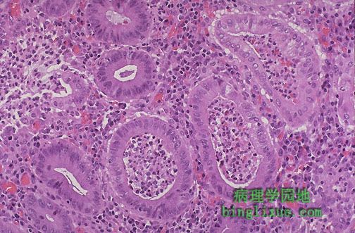 |
溃疡性结肠炎典型的组织学表现是粘膜隐窝小脓肿。不幸的是,并不是所有的炎症性肠病都能精确归类。 Crypt abscesses are a histologic finding more typical with ulcerative colitis. Unfortunately, not all cases of inflammatory bowel disease can be classified completely in all patients. |
 |
随着时间的推移,溃疡性结肠炎病人有患腺癌的危险。左侧可见很多正常腺体,但是右侧腺体明显异常,是第一个恶变的标志。 Over time, there is a risk for adenocarcinoma with ulcerative colitis. Here, more normal glands are seen at the left, but the glands at the right demonstrate dysplasia, the first indication that there is a move towards neoplasia. |