
 |
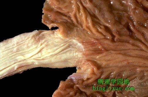
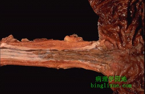
This is a normal esophagus with the usual white to tan smooth mucosa seen at the left. The gastroesophageal junction (not an anatomic sphincter) is at the center, and the stomach is at the right. 图左边示正常食管的浅褐色平滑黏膜。图中示胃-食管交界处( 并非解剖学括约肌位置)。图右边为胃部。 |
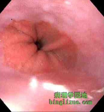 |
The upper GI endoscopic view of the transition from tan squamous mucosa to pink columnar mucosa is seen below. 上消化道胃镜所见到的由褐色的食管黏膜向胃部粉红色柱状黏膜过渡。 |
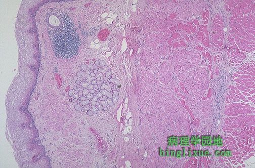 |
This is normal esophageal squamous mucosa at the left, with underlying submucosa containing mucus glands and a duct surrounded by lymphoid tissue. The muscularis is at the right. 图左边示正常食管鳞状上皮黏膜,黏膜下层内粘液腺体, 以及由淋巴组织包围的腺体导管。图右侧示食管肌层。 |
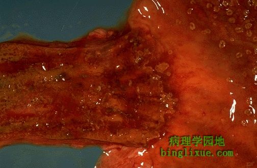 |
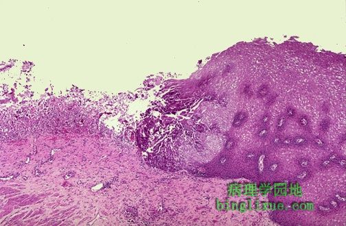
这是念球菌属食管炎。食管下段可见黄褐色的斑块,同时黏膜充血。右上部的胃也有同样的病变。 This is Candida esophagitis. Tan-yellow plaques are seen in the lower esophagus, along with mucosal hyperemia. The same lesions are also seen at the upper right in the stomach. |
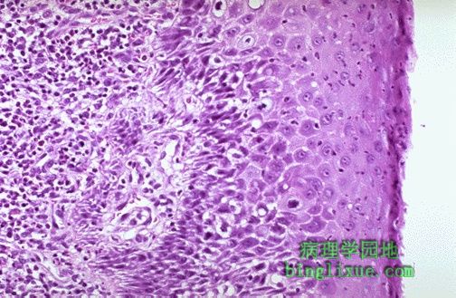 |
图示急性食管炎,黏膜下层中性粒细胞增多,中性粒细胞也浸润到右边的鳞状上皮层。 Acute esophagitis is manifested here by increased neutrophils in the submucosa as well as neutrophils infiltrating into the squamous mucosa at the right. |
 |
食管下段显示有边界清楚的溃疡,与左边正常的苍白色食管黏膜相比较,溃疡基底部呈红褐色。此种“穿孔”性溃疡提示有单纯疱疹病毒的感染。 The lower esophagus here shows sharply demarcated ulcerations that have a brown-red base, contrasted with the normal pale white esophageal mucosa at the far left. Such "punched out" ulcers are suggestive of herpes simplex infection. |
 |
疱疹性溃疡有明显的界限。左边溃疡基底面缺少鳞状上皮仅存有坏死的碎片。病变活检显示,鳞状上皮细胞核内包涵体提示为单纯疱疹病毒性食管炎。该病人由于化疗产生了免疫抑制。 A herpetic ulcer is seen microscopically to have a sharp margin. The ulcer base at the left shows loss of overlying squamous epithelium with only necrotic debris remaining. Biopsies of these lesions reveals intranuclear inclusions in squamous epithelial cells indicative of herpes simplex virus esophagitis. This patient was immune compromised from chemotherapy. |
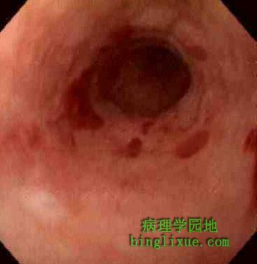 |
胃镜检查图,食管下部有圆的的溃疡红斑。 In the upper GI endoscopic view below, there are rounded, erythematous ulcerations of the lower esophagus. |
 |
高倍镜下,疱疹性溃疡边缘的鳞状上皮细胞内有浅粉红色的毛玻璃样物。一些内含物簇积在一起即多核化是另一个常见的病毒性细胞病理效应。 At high magnification, the squamous mucosa at the margin of the herpetic ulcer shows pale pink "ground glass" inclusions within squamous epithelial cells. Some of the inclusions are clustered together-- multinucleation is another common viral cytopathic effect. |
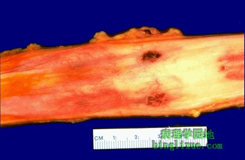 |
单纯疱疹病毒感染的免疫抑制病人,食管中部有两个边界明显的穿孔性溃疡。 Here are two more sharply demarcated "punched out" ulcerations of the mid esophagus in an immunocompromised patient with herpes simplex infection. |