
 |
在视野中心是一个嗜碱性细胞,它有一个多叶核(象PMN的核),并且细胞质里有为数众多的粗糙的、深蓝色的颗粒。它们在正常的外周血涂片中是很少见的,而且它们的意义还不确定。左上是一杆状核嗜中性粒细胞,右下可见一个大的、被激活的淋巴细胞。) There is a basophil in the center of the field which has a lobed nucleus (like PMN's) and numerous coarse, dark blue granules in the cytoplasm. They are infrequent in a normal peripheral blood smear, and their significance is uncertain. A band neutrophil is seen on the left, and a large, activated lymphocyte on the right. |
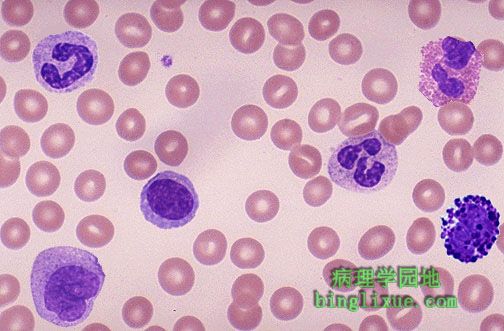 |
可见分叶核嗜中性粒细胞(左上),杆状核嗜中性粒细胞(中央偏右),淋巴细胞(中央偏左),单核细胞(左下),嗜酸性粒细胞(右上),嗜碱性粒细胞(右下)和血小板(杆状核嗜中性粒细胞右侧的小蓝块)。 Identify the segmented neutrophil, band neutrophil, lymphocyte, monocyte, eosinophil, basophil, and platelet. |
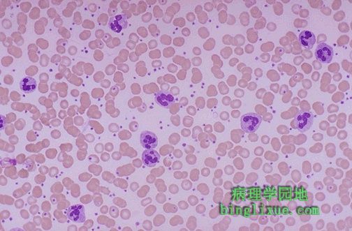 |
背景上的RBC是正常的,但有大量嗜中性粒细胞存在,主要因嗜中性粒细胞的增多而使WBC(白细胞)计数增高,提示有炎症或感染的存在。类白血病反应(非白血病)中,WBC(白细胞)计数非常高(>50000)。这种反应因嗜中性粒细胞中碱性磷酸酶(LAP)的大量存在而和恶性的白血病相区别。 The RBC's in the background appear normal. The important finding here is the presence of many PMN's. An elevated WBC count with mainly neutrophils suggests inflammation or infection. A very high WBC count (>50,000) that is not a leukemia is known as a "leukemoid reaction". This reaction can be distinguished from malignant WBC's by the presence of large amounts of leukocyte alkaline phosphatase (LAP) in the normal neutrophils. |
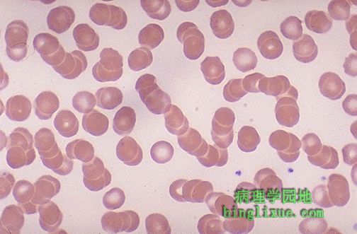 |
RBC成叠连状,称为“缗钱壮红细胞”,当血清蛋白质,特别是纤维蛋白原和球蛋白增加时出现。这样长的RBC链更容易沉积下来,这就是沉降率升高的机制,见于非特异性炎症时,急性期血清蛋白质增加。 The RBC's here have stacked together in long chains. This is known as "rouleaux formation" and it happens with increased serum proteins, particularly fibrinogen and globulins. Such long chains of RBC's sediment more readily. This is the mechanism for the sedimentation rate, which increases non-specifically with inflammation and increased "acute phase" serum proteins. |
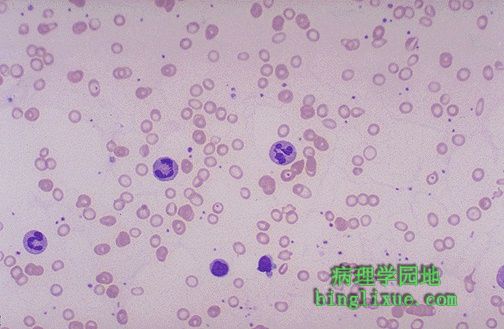 |
图示红细胞(RBC)与正常相比,体积变小且中心苍白区增加。提示有小细胞低色素性贫血。另外,红细胞大小不等形状多样。 The RBC's here are smaller than normal and have an increased zone of central pallor. This is indicative of a hypochromic (less hemoglobin in each RBC) microcytic (smaller size of each RBC) anemia. There is also increased anisocytosis (variation in size) and poikilocytosis (variation in shape). |
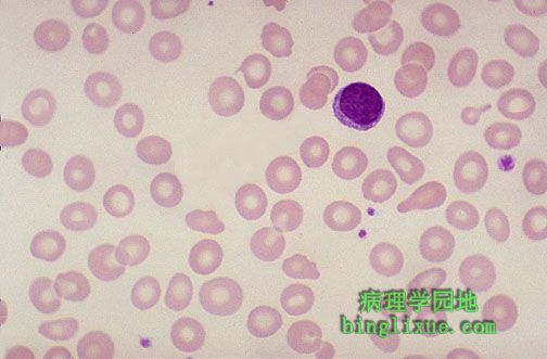 |
小细胞低色素性贫血最主要的原因是铁缺乏。最常见的营养缺乏是膳食中铁的不足。因此缺铁性贫血就较为常见。儿童和生育期妇女(从月经失血和妊娠期)是最危险人群。 The most common cause for a hypochromic microcytic anemia is iron deficiency. The most common nutritional deficiency is lack of dietary iron. Thus, iron deficiency anemia is common. Persons most at risk are children and women in reproductive years (from menstrual blood loss and from pregnancy). |
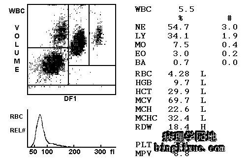 |
缺铁性贫血的CBC(完全血细胞计数)。可见血红蛋白(HGB)减少,MCV(红细胞平均体积)减少提示小细胞低色素性贫血。这里血红蛋白过少与MCH(红细胞平均血红蛋白)降低有关。 Here is data from a CBC in a person with iron deficiency anemia. Note the low hemoglobin (HGB). Microcytosis is indicated by the low MCV (mean corpuscular volume). Hypochromia correlates here with the low MCH (mean corpuscular hemoglobin). |
 |
巨幼细胞贫血(大细胞性贫血)中多叶核嗜中性粒细胞,有8叶,而不是正常的3到4叶。 Here is a hypersegmented neutrophil that is present with megaloblastic anemias. There are 8 lobes instead of the usual 3 or 4. Such anemias can be due to folate or to B12 deficiency. The size of the RBC's is also increased (macrocytosis, which is hard to appreciate in a blood smear). |
 |
恶性贫血,有巨卵形红细胞、多叶核嗜中性粒细胞。请比较红细胞与中央左下方的淋巴细胞的大小。 This hypersegmented neutrophil is present along with macro-ovalocytes in a case of pernicious anemia. Compare the size of the RBC's to the lymphocyte at the lower left center. |
 |
全血细胞计数显示MCV(红细胞平均体积)明显地升高,是典型的巨幼细胞贫血的表现。失血或溶血性贫血恢复期,MCV可略微升高。这是因为新释放的红细胞,网织红细胞,比正常红细胞体积大。而当其成熟后,体积会轻微减小。 The CBC here shows a markedly increased MCV, typical for megaloblastic anemia. The MCV can be mildly increased in persons recovering from blood loss or hemolytic anemia, because the newly released RBC's, the reticulocytes, are increased in size over normal RBC's, which decrease in size slightly with aging. |