 |
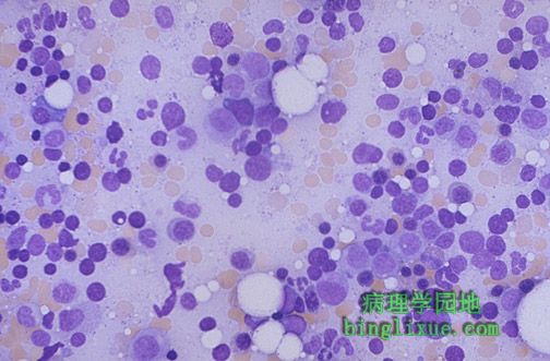
中倍镜正常骨髓的图象。可见巨核细胞,红细胞岛和粒细胞前体细胞。这是从一中年人的后髂嵴中取出来的骨髓,故约50%是细胞,并和骨髓成分混在一起。 This is the appearance of normal bone marrow at medium magnification. Note the presence of megakaryocytes, erythroid islands, and granulocytic precursors. This marrow is taken from the posterior iliac crest in a middle aged person, so it is about 50% cellular, with steatocytes admixed with the marrow elements. |
 |
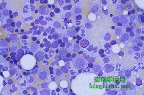
高倍镜下正常骨髓的图象。可见巨核细胞、红细胞岛和粒细胞前体细胞。这是从一中年人的后髂嵴中取出来的骨髓,故约50%是细胞,并和骨髓成分混在一起。 This is the appearance of normal bone marrow at high magnification. Note the presence of megakaryocytes, erythroid islands, and granulocytic precursors This marrow is taken from the posterior iliac crest in a middle aged person, so it is about 50% cellular, with steatocytes admixed with the marrow elements. |
 |
高倍镜下的正常骨髓涂片,可见红细胞前体细胞和粒细胞前体细胞。 This is the appearance of normal bone marrow smear at high magnification. Note the presence of erythroid precursors and granulocytic precursors. |
 |
高倍镜下的正常骨髓涂片,可见一个嗜酸性中幼粒细胞,一个嗜碱性中幼粒细胞和一个浆细胞。 This is the appearance of normal bone marrow smear at high magnification. Note the presence of an eosinophilic myelocyte, a basophilic myelocyte, and a plasma cell. |
 |
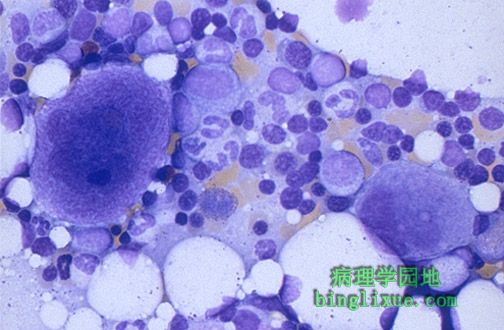
高倍镜下的正常骨髓涂片,可见巨核细胞,红细胞前体细胞和粒细胞前体细胞。 This is the appearance of normal bone marrow smear at high magnification. Note the presence of megakaryocytes, erythroid precursors, and granulocytic precursors. |
 |
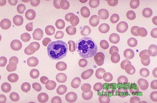
正常的、适中的红细胞形态,RBC中心大约1/3的区域是苍白的。RBC的形态可显示大小(红细胞大小不等症)和形状(异形红细胞症)上的最小的变异。同时可见少数模糊的蓝色的血小板。视野中心可见嗜中性粒细胞,左侧为杆状核,右侧为分叶状核。 The red blood cells here are normal, happy RBC's. They have a zone of central pallor about 1/3 the size of the RBC. The RBC's demonstrate minimal variation in size (anisocytosis) and shape (poikilocytosis). A few small fuzzy blue platelets are seen. In the center of the field are a band neutrophil on the left and a segmented neutrophil on the right. |
 |
自动化血细胞计数仪对正常的全血记数的资料,包括对WBC自动的分类计数。 This is normal data from a complete blood count as performed on an automated instrument, including an automated WBC differential count. |
 |
左侧可见一正常的、成熟的淋巴细胞,右侧是与其比较的分叶核嗜中性粒细胞。RBC是正常的淋巴细胞的2/3大小。 A normal mature lymphocyte is seen on the left compared to a segmented PMN on the right. An RBC is seen to be about 2/3 the size of a normal lymphocyte. |
 |
单核细胞稍微比淋巴细胞大,并有带褶皱的核。在细胞因子的影响下单核细胞从血流中迁移到组织中,成为巨噬细胞。在RBC之间可见很多小的污垢样的蓝色血小板。 Here is a monocyte. It is slightly larger than a lymphocyte and has a folded nucleus. Monocytes can migrate out of the bloodstream and become tissue macrophages under the influence of cytokines. Note the many small smudgy blue platelets between the RBC's. |
 |
视野中心可见一个两叶核嗜酸性粒细胞,并且细胞质里有众多的红色颗粒。在它的下方是一个小淋巴细胞。当过敏反应和寄生虫感染时嗜酸性粒细胞会增多。 In the center of the field is an eosinophil with a bilobed nucleus and numerous reddish granules in the cytoplasm. Just underneath it is a small lymphocyte. Eosinophils can increase with allergic reactions and with parasitic infestations. |