
 |
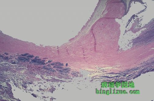
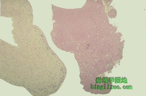
感染性心内膜炎瓣膜,左下方的蓝色菌落正向瓣膜的淡红色结缔组织蔓延。瓣膜血管较少,因此抗感染需要大剂量抗生素。 Here is a valve with infective endocarditis. The blue bacterial colonies on the lower left are extending into the pink connective tissue of the valve. Valves are relatively avascular, so high dose antibiotic therapy is needed to eradicate the infection. |
 |
感染性心内膜炎患者手指,甲下有小灶性出血,线性暗红色条纹状,此类出血也可见于外伤。 Seen here in the finger at the right are small splinter hemorrhages in a patient with infective endocarditis. These hemorrhages are subungual, linear, dark red streaks. Similar hemorrhages can also appear with trauma. |
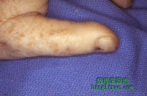 |
感染性心内膜炎患者左手大拇指的指甲下可见小线型灶状出血,血培养呈金黄*色葡萄球菌阳性。 Another small linear splinter hemorrhage is seen here subungually on the left thumb of a patient with infective endocarditis and blood culture positive for Staphylococcus aureus. |
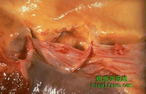 |
在最右方尖端边缘的小而淡红的赘生物是非细菌性血栓性心内膜炎(或称为“消耗性心内膜炎”)的典型表现;本病不是由感染引起的。它常见于血液为高凝固状态的人(Trousseau’s 综合症,一种与恶性肿瘤有关的副肿瘤综合征)以及重病者。 The small pink vegetation on the rightmost cusp margin represents the typical finding with non-bacterial thrombotic endocarditis (or so-called "marantic endocarditis"). This is non-infective. It tends to occur in persons with a hypercoagulable state (Trousseau's syndrome, a paraneoplastic syndrome associated with malignancies) and in very ill persons. |
 |
最左方尖端的消耗性赘生物,大小虽然很少超过0.5厘米,但是非常容易形成栓塞。 Here is another marantic vegetation on the leftmost cusp. These vegetations are rarely over 0.5 cm in size. However, they are very prone to embolize. |
 |
图示左侧为心脏瓣膜,右侧颜色较浅的为疣状赘生物。此赘生物呈现红色,是由纤维素和血小板构成的。此赘生物在形态学上显示像一个棕色的纸袋。此种淡粉色的赘生物是在非细菌性血栓性心内膜炎的时候形成的特征性改变。 The valve is seen on the left, and a bland vegetation is seen on the right. It appears pink because it is composed of fibrin and platelets. It displays about as much morphologic variation as a brown paper bag. Such bland vegetations are typical of the non-infective forms of endocarditis. |
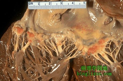 |
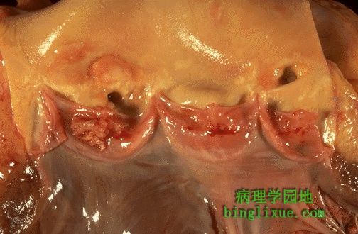
小疣状赘生物呈串珠状沿着心脏瓣膜的闭锁缘排列。本例病人患有系统性红斑狼疮,这类赘生物可见于任何瓣膜乃至心内膜上,并且在形态上与Libman-Sacks心内膜炎 的赘生物一致。系统性红斑狼疮病人仅有4%出现,并且赘生物小不易脱落,因此对机体的影响较小。注意腱索变厚、缩短、粘连,这是风湿性心脏病的表现。 Here are flat, pale tan, spreading vegetations over the mitral valve surface and even on the chordae tendineae. This patient has systemic lupus erythematosus. Thus, these vegetations that can be on any valve or even on endocardial surfaces are consistent with Libman-Sacks endocarditis. These vegetations appear in about 4% of SLE patients and rarely cause problems because they are not large and rarely embolize. Note also the thickened, shortened, and fused chordae tendineae that represent remote rheumatic heart disease. |
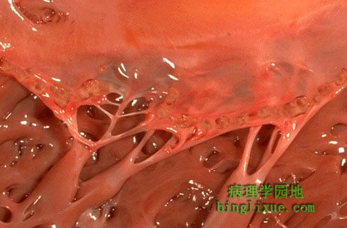 |
沿着二尖瓣的闭锁缘可见一些小的疣状赘生物,它们与急性风湿热相关。这些疣状赘生物平均仅几个毫米,沿着心内膜炎症区上的瓣膜闭锁缘形成。这种赘生物小不至于引起严重的心脏疾病。 The small verrucous vegetations seen along the closure line of this mitral valve are associated with acute rheumatic fever. These warty vegetations average only a few millimeters and form along the line of valve closure over areas of endocardial inflammation. Such verrucae are too small to cause serious cardiac problems. |
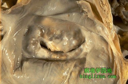 |
左心房可见二尖瓣,呈特有的鱼口状狭窄,并有慢性风湿病瘢痕。风湿性心脏病时二尖瓣最易受累,其次为二尖瓣和主动脉同时受累,然后是主动脉单独受累,二尖瓣、主动脉和三尖瓣同时受累最不常见。 The heart has been sectioned to reveal the mitral valve as seen from above in the left atrium. The mitral valve demonstrates the typical "fish mouth" shape with chronic rheumatic scarring. Mitral valve is most often affected with rheumatic heart disease, followed by mitral and aortic together, then aortic alone, then mitral, aortic, and tricuspid together. |
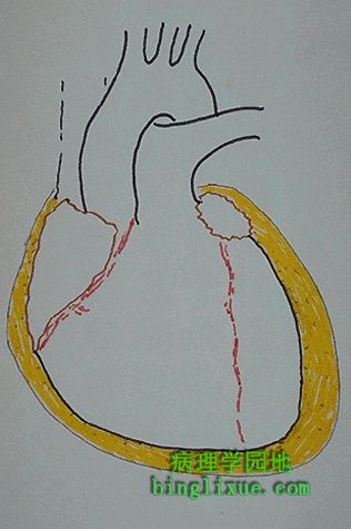 |
图示浆液性心包炎的形态。由于炎症较轻,因此未见纤维蛋白渗出。黄*色液体中以及心外膜表面上可见一些黑点,是零散的炎性细胞。浆液性心包炎以分泌液体聚集为特征,大量浆液渗出时就可能引起心包填塞。 This diagram depicts the appearance of a serous pericarditis. The amount of inflammation is minimal, so no exudation of fibrin occurs. The dark stippled dots in the yellow fluid and on the epicardial surface represent scattered inflammatory cells. Serous pericarditis is marked by fluid collection. Rarely, the fluid collection may be large enough to cause tamponade. |