
 |
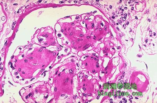
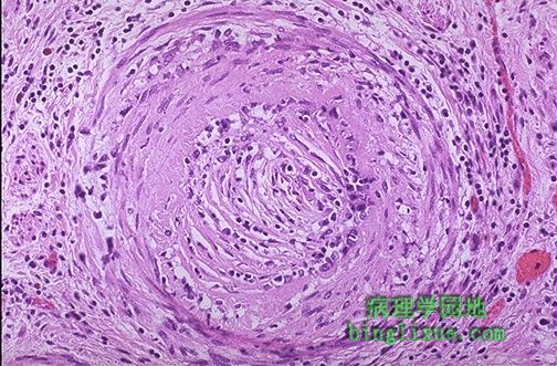
除动脉粥样硬化之外,还有其它两种形式的动脉硬化(动脉变硬)∶细动脉硬化症和动脉中层钙化。肾脏可见典型的细动脉硬化。经过PAS染色,可见连接右下方肾小球的细动脉已明显变厚,它就是被称为玻璃样变细动脉硬化的一种形态。玻璃样变细动脉硬化可见于老年人,但是在有糖尿病和/或有高血压的病人中有着更严重的病变。 There are two other forms of arteriosclerosis (hardening of the arteries) in addition to atherosclerosis: arteriolosclerosis and medial calcific sclerosis. Arteriolosclerosis is typically seen in the kidneys. One form, called hyaline arteriolosclerosis, is demonstrated by the markedly thickened arteriole to the lower right of this glomerulus with PAS stain. Hyaline arteriolosclerosis is seen in the elderly, but more advanced lesions are seen in persons with diabetes mellitus and/or with hypertension. |
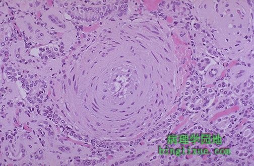 |
细动脉硬化的第二种形态。细动脉呈“洋葱皮”外观,这是增生性细动脉硬化的典型表现。这种病变常常与恶性高血压有关。 The second form of arteriolosclerosis is shown here. The arteriole here has an "onion skin" appearance typical of hyperplastic arteriolosclerosis. This lesion is most often associated with malignant hypertension. |
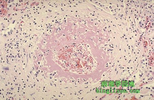 |
肾脏增生性细动脉硬化合并高血压,如图发生纤维素样坏死。 One complication of hyperplastic arteriolosclerosis with malignant hypertension is fibrinoid necrosis, as seen here in a renal arteriole. |
 |
甲状腺组织右侧肌性动脉可见Monckeberg氏中层钙化。它最常见于老年人,但临床表现不明显。钙化动脉还可能在拍X线照片时显现,从而发现它的形态。钙沉积在肌性动脉的中层,特别是在骨盆和颈部的动脉。 Monckeberg's medial calcific sclerosis is seen in this artery to the right of thyroid tissue at the left. This finding occurs most often in the elderly and is of no clinical significance, other than that the calcified arteries may be visualized on radiographs, and you need to know what is represented. Calcium deposits collect in the media of muscular arteries, particularly in pelvis and neck. |
 |
冠状动脉影响到心脏移植病人能否长期存活,它的特征有内膜增生伴有心外膜以及心肌内的冠状动脉分支管腔狭窄,从而导致缺血性改变。 A rate-limiting step to long-term survival in heart transplant patients is coronary arteriopathy, which is marked intimal proliferation with lumenal narrowing of the small epicardial and intramyocardial coronary artery branches, leading to ischemic changes. |
 |
肌性动脉有慢性炎性细胞浸润的血管炎存在。可见血管内皮细胞增生以及管腔消失。通常,血管炎是自身免疫性疾病的特征性表现,如本例病人就患有系统性红斑狼疮。血管炎是罕见的而且它形态的多样性容易造成混淆,从而难以诊断和区分。测试抗核抗体和抗中性粒细胞抗体可帮助诊断。 This muscular artery demonstrates vasculitis with chronic inflammatory cell infiltrates. The endothelial cells have proliferated and the lumen is absent. Often, vasculitis is a feature of an autoimmune disease, such as systemic lupus erythematosus as was present in this patient. Vasculitis is uncommon and the various forms are confusing and difficult to diagnosis and classify. Tests such as ANA and ANCA help. |
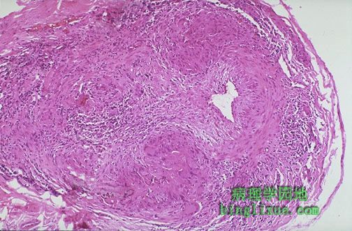 |
颞动脉炎是巨细胞性动脉炎的一种,巨细胞性动脉炎主要影响颈外动脉的分支,但有时也会影响主动脉弓和冠状动脉。在血管中膜可发现灶性肉芽肿。 Temporal arteritis is one manifestation of giant cell arteritis, which can affect mainly branches of external carotid artery, but sometimes also the great vessels at the aortic arch and coronaries. There is focal granulomatous inflammation of the media. |
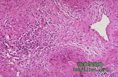 |
巨细胞动脉炎(颞动脉)在50岁以前较少见。血沉速率常常明显地升高(可达100 mm/hr甚至更高)。半数以上的病人有风湿性多肌痛。高倍镜下可见病灶的肉芽肿炎症伴有动脉腔狭窄。 Giant cell (temporal) arteritis is uncommon before age 50. The sedimentation rate is often markedly elevated (100 mm/hr or more). Half of patients have polymyalgia rheumatica. The focal granulomatous inflammation with narrowed arterial lumen is seen here at high magnification. |
 |
在巨细胞性动脉炎患者头皮的表面可能见到坚硬有触痛的颞动脉。 Patients with giant cell arteritis may have a visible firm, palpable, painful temporal artery that courses over the surface of the scalp. |
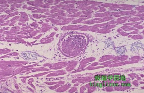 |
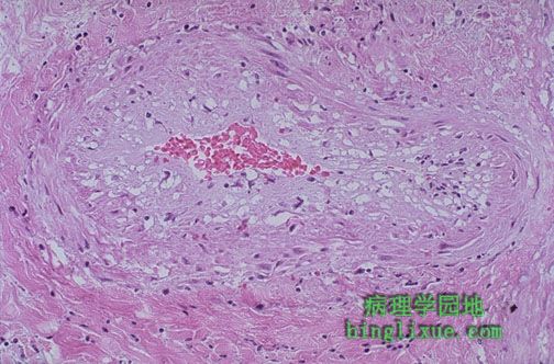
血栓性血小板减少性紫癜的特征性表现是心脏外周小动脉形成由纤维蛋白和血小板组成血栓的形成。因此,有血栓性血小板减少性紫癜的患者可能死于心力衰竭。 This fibrin and platelet thrombus in a small peripheral artery in the heart is characteristic for thrombotic thrombocytopenic purpura. For this reason, patients with TTP may die from heart failure. |