
 |
This high magnification of the atheroma shows numerous foam cells and an occasional cholesterol cleft. A few dark blue inflammatory cells are scattered within the atheroma. 图示:动脉粥样硬化高倍镜显示多量泡沫细胞,偶见胆固醇结晶(棱状空隙)。一些暗蓝色的炎细胞散在分布于粥样硬化病灶内。 |
 |
This is about as normal as an adult aorta in America gets. The faint reddish staining is from hemoglobin that leaked from RBC's following death. The surface is quite smooth, with only occasional faint small yellow lipid streaks visible. 这是美国正常成年人的大动脉。暗红色的污点是红细胞坏死后漏出的血色素造成的。表面非常光滑,偶尔见到微小的黄*色脂质条纹。 |
 |
Put down that jelly doughnut and look carefully at this aorta. The white arrow denotes the most prominent fatty streak in the photo, but there are other fatty streaks scattered over the aortic surface. Fatty streaks are the earliest lesions seen with atherosclerosis in arteries. 放下手头上的东西,认真观察这张大动脉图片。图中白色箭头指示的是最为明显的脂质条纹,同时也有一些散在分布的脂质条纹。脂纹是动脉粥样硬化早期的损伤性表现。 |
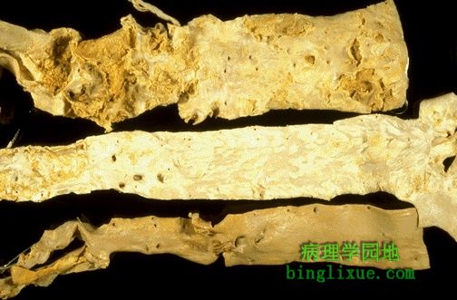 |
These three aortas demonstrate mild, moderate, and severe atherosclerosis from bottom to top. At the bottom, the mild atherosclerosis shows only scattered lipid plaques. The aorta in the middle shows many more larger plaques. The severe atherosclerosis in the aorta at the top shows extensive ulceration in the plaques. 从下到上依次排列着轻、中、重三种不同程度的动脉粥样硬化。下面轻度粥样硬化仅见散在脂斑。中间的大动脉显示更多、更大的斑块。上面最重的粥样硬化动脉显示在斑块上有大量溃疡形成。 |
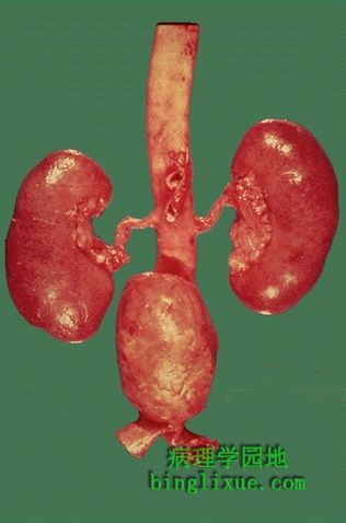 |
Here is an example of an atherosclerotic aneurysm of the aorta in which a large "bulge" appears just above the aortic bifurcation.Such aneurysms are prone to rupture when they reach about 6 to 7 cm in size. They may be felt on physical examination as a pulsatile mass in the abdomen.Most such aneurysms are conveniently located below the renal arteries so that surgical resection can be performed with placement of a dacron graft. 图示:动脉 分支的上方有个大动脉粥样硬化形成的动脉瘤。当这种动脉瘤达到6-7cm时就很容易破裂。体格检查可在腹部查到搏动的肿块。大多这样的动脉瘤位于肾动脉下面附近,这就使得临床外科手术切除得以更好的执行,原因是在此处可以有效进行人工修补。 |
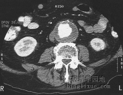 |
This abdominal high speed CT scan with contrast demonstrates an abdominal aortic aneurysm approximately 6 cm in diameter. At this size, there is increased risk for rupture. 高速CT腹部扫描显示腹主动脉直径约6cm,它的破裂风险很大。 |
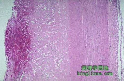 |
This microscopic cross section of the aorta shows a large overlying atheroma on the left. Cholesterol clefts are numerous in this atheroma. The surface on the far left shows ulceration and hemorrhage. Despite this ulceration, atheromatous emboli are rare (or at least, complications of them are rare). 动脉纵切镜下显示左侧有大的动脉粥样硬化病灶。有大量的胆固醇结晶。最左面显示了溃疡和出血。即使这样溃疡和血栓形成是少见的,或者说动脉粥样硬化的复合病变是少见的。 |
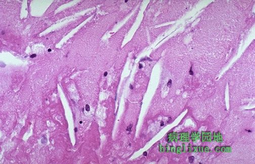 |
This is a high magnification of the aortic atheroma with foam cells and cholesterol clefts. 高倍显示动脉粥样硬化时的胆固醇结晶和泡沫细胞。 |
 |
This is severe atherosclerosis of the aorta in which the atheromatous plaques have undergone ulceration along with formation of overlying mural thrombus. 严重的动脉粥样硬化病灶内形成了溃疡同时也伴有血管壁上血栓的形成。 |
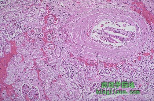 |
Despite the frequency of aortic atherosclerosis, cholesterol emboli are rare, or at least insignficant most of the time. Seen here in a renal artery branch are cholesterol clefts of such an embolus. This patient had severe ulcerative, friable atheromatous plaques and had undergone angiography, which increases the risk for such emboli. 虽然动脉粥样硬化比较常见,但是胆固醇栓子是少见的,至少是大多情况下不明显的。图示肾动脉分形成了这种胆固醇栓子。该病人有严重的溃疡形成,易碎的粥样斑块,同时也做了会增加血栓形成危险的血管造影术。 |