
 |
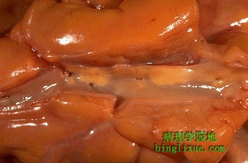
冠状动脉被纵向剖开。冠状动脉从左至右通过照片的中间,周围围绕着心外膜脂肪。心外膜脂肪量增加与肥胖有关。大量脂肪是动脉粥样硬化的危险因素。此处仅见轻度的动脉粥样硬化,同时仅有一暂时性的黄褐色脂质斑块但未见管腔狭窄。 A coronary artery has been opened longitudinally. The coronary extends from left to right across the middle of the picture and is surrounded by epicardial fat. Increased epicardial fat correlates with increasing total body fat. There is a lot of fat here, suggesting one risk factor for atherosclerosis. This coronary shows only mild atherosclerosis, with only an occasional yellow-tan lipid plaque and no narrowing. |
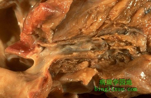 |
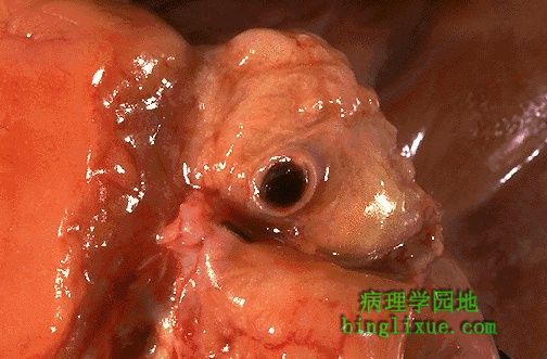
源于主动脉根左边的左冠状动脉。横跨照片中部延至右侧的是左冠状动脉前降支。可见重度的动脉粥样硬化伴随大片的钙化。右侧较远处、可见一明显狭窄。 This is the left coronary artery from the aortic root on the left. Extending across the middle of the picture to the right is the anterior descending branch. This coronary shows severe atherosclerosis with extensive calcification. At the far right, there is an area of significant narrowing. |
 |
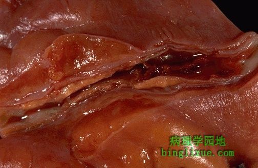
图片中央显示冠状动脉粥样硬化合并粥样斑块内出血,出血使得动脉管腔明显狭窄。 This is coronary atherosclerosis with the complication of hemorrhage into atheromatous plaque, seen here in the center of the photograph. Such hemorrhage acutely may narrow the arterial lumen. |
 |
见冠状动脉前降支的横切面有明显粥样硬化及管腔狭窄,左边动脉的近端部分尤为明显(自左向右依次排列着从近端到远端的切面)。通常粥样硬化最严重的是近端,因为近端血流形成更多湍流。病灶越多血管成形术或分流术的操作越复杂。 Cross sections of this anterior descending coronary artery demonstrate marked atherosclerosis with narrowing. This is most pronounced at the left in the more proximal portion of this artery. In general, the worst atherosclerosis is proximal, where arterial blood flow is more turbulent. More focal lesions mean that angioplasty or bypass can be more useful procedures. |
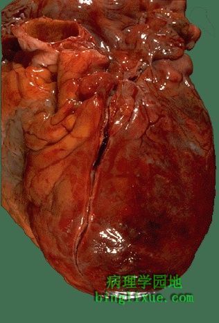 |
心脏的前表面可见一敞口的冠状动脉左前降支。管腔内见一暗红色新鲜血栓,血栓右下方位于闪亮心外膜之下的心肌呈暗红色与梗死后的颜色一致。 The anterior surface of the heart demonstrates an opened left anterior descending coronary artery.Within the lumen of the coronary can be seen a dark red recent coronary thrombosis. The dull red color to the myocardium as seen below the glistening epicardium to the lower right of the thrombus is consistent with underlying myocardial infarction. |
 |
放大后可见冠状动脉管腔内的暗红色血栓较明显,粥瘤的黄褐色斑块使得冠状动脉明显狭窄,并且血栓完全阻塞了管腔。 At high magnification, the dark red thrombus is apparent in the lumen of the coronary. The yellow tan plaques of atheroma narrow this coronary significantly, and the thrombus occludes it completely. |
 |
此横截面显示冠状动脉血栓形成。血栓形成严重阻碍了血流并导致局部缺血和/或梗死,临床出现急剧胸痛。 A thrombosis of a coronary artery is shown here in cross section. This acute thrombosis diminishes blood flow and leads to ischemia and/or infarction, marked clinically by the sudden onset of chest pain. |
 |
左心室壁纵切面可见一大块新近的心肌梗死区,其中坏死心肌呈黄褐色,周围是红色的充血区,其余的正常心肌呈红褐色。 This is the left ventricular wall which has been sectioned lengthwise to reveal a large recent myocardial infarction. The center of the infarct contains necrotic muscle that appears yellow-tan. Surrounding this is a zone of red hyperemia. Remaining viable myocardium is reddish- brown. |
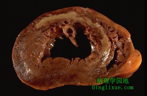 |
心脏横截面显示左心室在左边。从前部直到室间隔是一大片新近梗死区,围绕中部的充血区呈棕褐色。梗死是透壁性的即累及心肌的全层。 This cross section through the heart demonstrates the left ventricle on the left. Extending from the anterior portion and into the septum is a large recent myocardial infarction. The center is tan with surrounding hyperemia. The infarction is "transmural" in that it extends through the full thickness of the wall. |
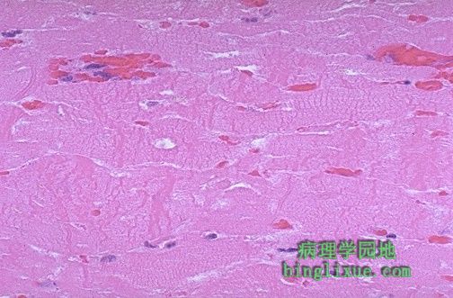 |
急性心肌梗死第一天组织学上表现为明显的收缩带坏死。在大多数可见细胞中,心肌纤维的横纹丢失,细胞核模糊。注意在纤维上有许多无规律暗红的呈波浪状的收缩带经过。 The earliest change histologically seen with acute myocardial infarction in the first day is contraction band necrosis. The myocardial fibers are beginning to lose cross striations and the nuclei are not clearly visible in most of the cells seen here. Note the many irregular darker pink wavy contraction bands extending across the fibers. |