
 |
病理学上,SLE患者皮肤可表现为脉管炎和真皮慢性炎性浸润,如图所示。自身免疫疾病患者,可在许多不同的器官发生脉管炎,且容易与风湿病的症状和体征相混淆. Histologically, the skin in SLE may demonstrate a vasculitis and dermal chronic inflammatory infiltrates, as seen here. Vasculitis with autoimmune disease can occur in many different organs and can lead to the often confusing signs and symptoms of patients with rheumatic diseases. |
 |
图示SLE病人更严重的上皮层的炎性真皮浸润。在基底层已有空泡形成和溶解,在上皮层还有一个由红细胞形成的紫癜(皮疹形成的原因). Here is a more severe inflammatory skin infiltrate in the upper dermis of a patient with SLE in which the basal layer is undergoing vacuolization and dissolution, and there is purpura with RBC's in the upper dermis (which are the reason for the rash). |
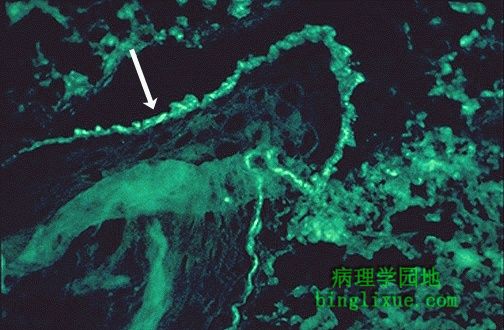 |
如果针对补体的抗体或免疫球蛋白进行免疫荧光染色,我们就能在表皮与真皮的交界处看到一条明亮荧光带,表明有免疫复合物的存在. |
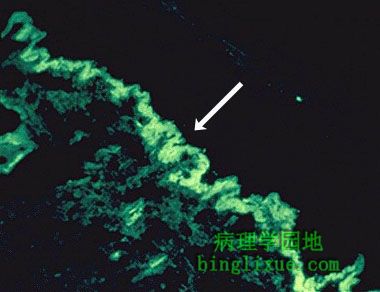 |
图示IgG抗体的免疫荧光,表明了免疫复合物在表皮与真皮的交界处.如果仅在有疹的皮肤上能看到的话,诊断可能是DLE.但是如果无疹的皮肤也是如此的话,那么诊断就是SLE. Here is another immunofluorescence staining pattern with antibody to IgG showing evidence for immune complexes at the dermal-epidermal junction. If such a pattern is seen only in skin involved by a rash, then the diagnosis is probably DLE, but if this pattern appears even in skin uninvolved by a rash, then the diagnosis is SLE. |
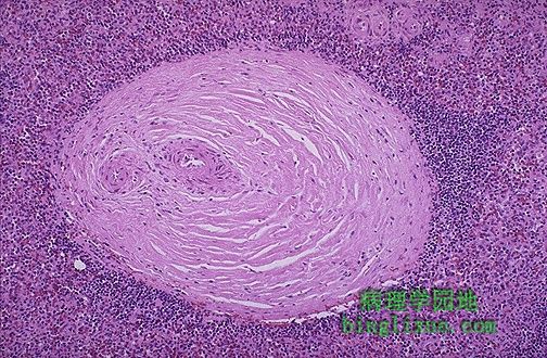 |
SLE病人尸检,可见其脾动脉外膜纤维化(“洋葱皮状”),尽管没有明显的临床症状。脉管炎导致了此结果。 The periarteriolar fibrosis ("onion skinning") seen in the spleen in patients with SLE at autopsy is quite striking, though of no major clinical consequence. This results from vasculitis. |
 |
风湿病较为严重的并发症之一是肾衰。肾衰更可能发生于SLE。图示肾小球,其中的毛细血管袢中呈明显的粉红色、管腔增厚,以至于毛细血管腔难以看见。即狼疮肾。 One of the feared complications of the rheumatic diseases is renal failure. This is most likely to occur with SLE. Here is a glomerulus in which the capillary loops are markedly pink and thickened such that capillary lumens are hard to see. This is lupus nephritis. |
 |
图示狼疮性肾炎病人肾小球,可见粉红色浓集的毛细血管袢,所谓“电线袢”,周围的肾小管不明显. Here is a glomerulus with thickened pink capillary loops, the so-called "wire loops", in a patient with lupus nephritis. The surrounding renal tubules are unremarkable. |
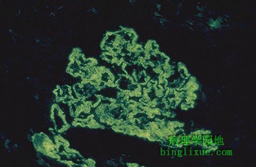 |
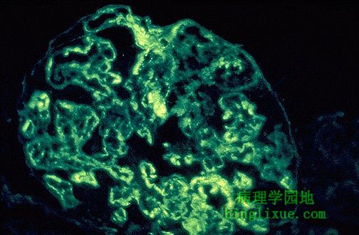
针对IgG抗体的免疫荧光染色,可见颗粒状荧光,表明免疫复合物沉积在肾小球毛细血管袢的基底膜上。
If immunofluorescence staining is performed, here with antibody to IgG, then a granular pattern of immunofluorescence is seen, indicative of deposition of immune complexes in the basement membranes of the glomerular capillary loops. |
 |
针对补体C1q的抗体的免疫荧光染色,肾小球可见颗粒状荧光,对SLE特异性强。 Here is another granular pattern of immunofluorescence in the glomerulus, this time with antibody to C1q complement, which is more specific for SLE. |
 |
电镜图示,由于免疫复合物沉积在肾小球毛细血管袢中,使基底膜增厚(箭头)。暗淡的免疫复合物主要位于内膜下。 The thickened basement membrane (arrow) that results from immune complex deposition in the glomerular capillary loop is prominent in this electron micrograph. The dark immune deposits are located mainly in a subendothelial position. |