|
|
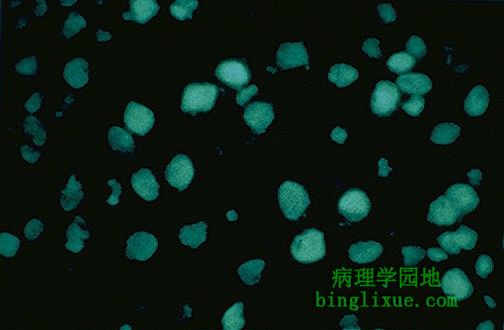
自身免疫性疾病的筛查常用抗核抗体(ANA)检测,病人血清与组织的底物温育,抗核抗体和相应的核抗原结合, 接着加入荧光抗体,在荧光显微镜下观察,看是否被染色。图示典型的抗核抗体染色阳性,呈均质型。
Screening for autoimmune diseases is often performed with the antinuclear antibody (ANA) test in which patient serum is incubated with a tissue substrate to which any autoantibodies to nuclear antigens will bind. Then, a fluorosceinated antibody is added and the tissue is observed under fluorescence microscopy to see if staining is present. Seen here is the typical "homogenous" pattern(均质型) of nuclear staining of a positive ANA.
|
|
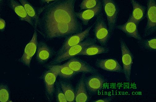
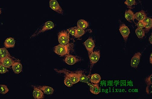
另例抗核抗体阳性,底物细胞是人类的Hep2. 任何报告都是抗核抗体阳性与效价同时报告的。效价是阳性病人血清简单稀释的结果.检测高于1:16 或 1:20 为阳性.通常效价越高越有可能存在着自身免疫性疾病.效价为1:40可能并不意味着什么,但1:1012的效价当然就不能被忽视了。
Here is another positive ANA in which the substrate cells are a human cell line known as Hep2. Any positive ANA is reported with a titer. The titer is simply the dilution of patient serum at which the test is still positive. The test has to be positive at greater than 1:16 or 1:20 (depending upon the lab) to be positive at all. In general, the higher the titer, the more likely a serious autoimmune disease is present. Thus, a 1:40 result might not mean much, but a 1:1012 certainly should not be ignored.
|
|
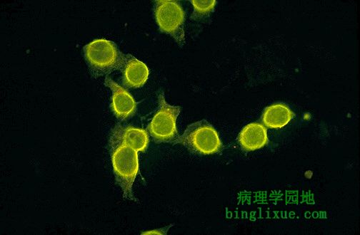
抗核抗体检测时底物细胞显出特殊的染色模式.图示:核膜型(周边型),是大多数SLE患者具有的特点.
Sometimes when performing the ANA test, the substrate cells demonstrate particular patterns of staining. This is the so-called "rim" pattern that is more characteristic of SLE.
|
|
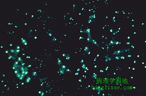
短膜虫属原虫的唯一目的是作为双链DNA检测的底物.图示含有明亮的荧光动力体(在特定的原生物中位于鞭毛某部附近的独立复制的细胞结构)
是阳性结果的标志.双链DNA检测结果阳性则强烈提示SLE.
These are Crithidia organisms, whose sole purpose for existence is to serve as a substrate for the double stranded DNA test. Here the little critters have brightly fluorescing kinetoplasts indicative of a positive test. A positive double stranded DNA test strongly suggests a diagnosis of SLE.
|
|
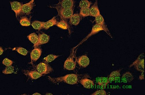
图示斑点型(又称核颗粒型、核斑块型),是针对可提取的核抗原,特别是核蛋白,的自身抗体的典型显现方式.此型并不是很特异,但可见于混合结缔组织病,包括SLE,硬皮病和多发性肌炎,但不伴有严重肾脏或肺脏疾病.甚至对于专家来说,自身免疫性疾病很难分类。
This is the so-called "speckled" pattern of staining which is more characteristic of the presence of autoantibodies to extractable nuclear antigens, particularly ribonucleoprotein. This pattern is not very specific, but may be seen with an entity called "mixed connective tissue disease" which is a mix between SLE, scleroderma, and polymyositis, but without serious renal or pulmonary disease. The autoimmune diseases are very hard to classify, even for the experts.
|
|
图示核仁型,在HEP2细胞核仁中,可见明亮的荧光.此型进一步提示进行性系统性硬化病。
This is the so-called "nucleolar pattern" of staining in which the bright fluorescence is seen within the nucleoli of the Hep2 cells. This pattern is more suggestive of progressive systemic sclerosis.
|
|
另例核仁型。更象从哈伯望远镜观察到的景象。
Here is another nucleolar pattern of staining that is more reminiscent of a view from the Hubble space telescope (before the repair).
|
|
图示著名的“LE细胞”实验,它的价值仅仅在于表明自身抗体的作用机理。粉红色斑点是变性的细胞核.图中可见两个,一个被嗜中性粒细胞吞噬.该实验没有抗核抗体检测敏感,因此抗核抗体检测取代了LE细胞实验.现在不要求作LE细胞检测.
Here is the famous "LE cell" test which has value only in demonstrating how the concept of autoantibodies work. The pink blobs are denatured nuclei. Here are two, with one seen being phagocytozed in the center by a PMN. This test is not nearly as sensitive as the ANA which has supplanted the LE cell test. Therefore, NEVER order an LE cell test.
|
|
颊疹(又叫蝴蝶斑,因其位于两颊呈蝴蝶状)提示狼疮.盘状红斑狼疮(DLE)与(系统性红斑狼疮)SLE相比相对轻些,主要表现在皮肤上。两者暴晒于阳光下都可以加速红斑的产生.一小部分DLE患者大约(5-10%)发展成SLE.(通常DLE 患者抗核抗体为阳性).
The young woman has a malar rash (the so-called "butterfly" rash because of the shape across the cheeks). Such a rash suggests lupus. Discoid lupus erythematosus (DLE) involves mainly just the skin and is, therefore, relatively benign compared to systemic lupus erythematosus (SLE). In either case, sunlight exposure accentuates this erythematous rash. A small number (5 to 10%) of DLE patients go on to develop SLE (usually the DLE patients with a positive ANA).
|
|
图示典型颊疹,狼疮比其它自身免疫性疾病的颊疹更为典型,但不是最特异的。
Here is a more subtle malar rash. Such a rash is more typical for lupus than other autoimmune diseasease, but is not entirely specific for it.
|