|
|
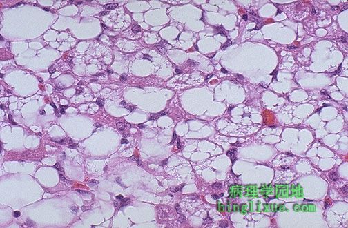
At high magnification, large bizarre lipoblasts are seen in this liposarcoma. Sarcomas are best treated surgically, because most respond poorly to chemotherapy or radiation.
图示:脂肪肉瘤
高倍镜下可见大而畸形的成脂细胞。
最好的治疗方法是外科切除,因为对化疗和放疗不敏感。 |
|
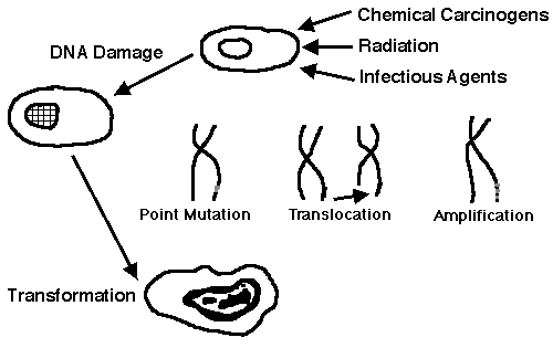
The process of neoplasia begins with cell transformation. A variety of chemical carcinogens as diverse as benzene, cigarette smoke, and nitrites can initiate and/or promote this process. Radiation, either as low level long-term environmental gamma rays or as higher dose therapeutic radiation, can also produce genetic mutations. Infectious agents such as human papillomavirus can lead to cellular transformation as well.
Genetic damage with DNA alterations leads to point mutations of genes, translocations of genetic material between chromosomes, and gene reduplication with amplification. These alterations transform proto-oncogenes into oncogenes. The proto-oncogenes may play a role in growth promotion and regulation in normal cells, perhaps in embryogenesis, but are typically "turned off" in adults. They are "turned on" by transformation.
肿瘤形成源于细胞转化。多种多样的化学致癌物如苯、吸烟、亚硝酸盐都能够引起(和)或促进肿瘤的发生。不管是周围环境中低水平长期的γ射线的作用,还是高剂量的放疗都会导致基因突变。感染性因素如人类乳头状瘤病毒能导致细胞转化。
伴随DNA改变的基因损伤会导致基因的点突变、染色体间基因移位和扩增性基因复制。这些改变能促使原癌基因改变为癌基因。原癌基因或许在胚胎期正常细胞的增生和调节起着非常重要的作用,然而成年人原癌基因处于“关闭”状态,但是通过转化可以使其启动。 |
|
Neoplasia, or uncontrolled cellular proliferation, can result either from mutations that "turn on" the oncogenes that stimulate growth, or from mutations that result in loss of tumor suppressor genes and their products that inhibit growth.
肿瘤或失去控制的细胞增生是由于突变使癌基因启动从而促使增生,或者由于肿瘤抑制基因和肿瘤抑制基因产物的缺失。 |
|
|
|
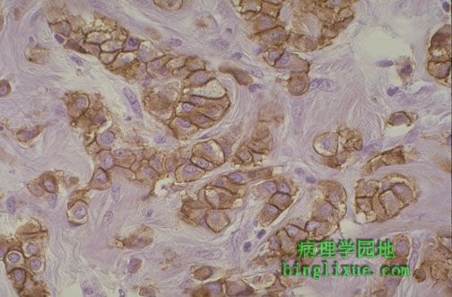
Here is an example of c-erb-B2 positivity in a breast carcinoma. This oncogene acts via reduplication of the normal proto-oncogene hundreds of times, leading to production of a protein product that drives unregulated cell growth. This is detected here by immunoperoxidase staining with the brown reaction product concentrated in a perimembranous pattern around the cells.
图示:乳腺癌c-erb-B2呈阳性
此癌基因通过使正常原癌基因数百次的复制产生一种能驱使细胞非调节性生长的蛋白。
免疫过氧化物酶染色显示在细胞周围可见褐色的膜状物。 |
|
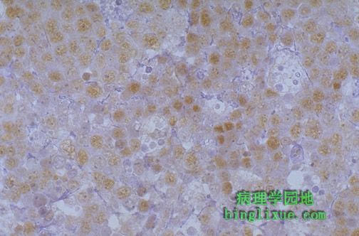
This is an example of c-myc positivity in a carcinoma. This oncogene acts via DNA transcriptional activation. The nuclear binding is demonstrated here by immunoperoxidase staining in which the brown reaction product is localized to nuclei.
图示:c-myc阳性
此癌基因能使DNA翻译激活。免疫过氧化物酶染色显示细胞核呈褐色。 |
|
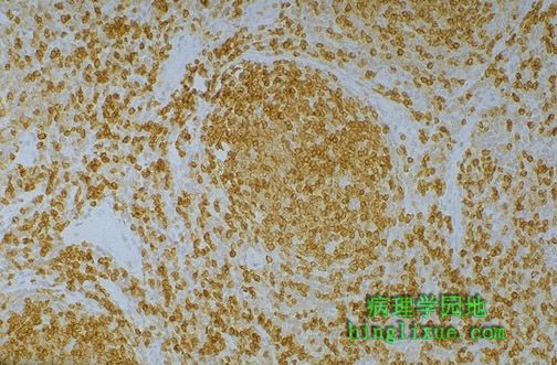
This is an example of bcl-2 positivity in a lymphoma. In this case, the overexpression of this oncogene results in an inhibition of apoptosis, and increased numbers of lymphocytes. The immunoperoxidase stain here highlights the lymphocytes in lymphoid follicles and interfollicular areas.
图示:淋巴瘤bcl-2阳性
此种基因的过度表达抑制了细胞凋亡,淋巴细胞数量增多。
免疫过氧化物酶染色显示集中在淋巴滤泡内和淋巴滤泡之间。 |
|
|
Staging and grading schema have been devised for malignant neoplasms, because the stage and/or grade may determine the treatment and the prognosis. In general, the higher the stage, the larger a neoplasm is and the farther it has likely spread.
Staging
The most common systems for staging employs the TNM classification. A "T" score is based upon the size and/or extent of invasion. The "N" score indicates the extent of lymph node involvement. The "M" score indicates whether distant metastases are present. Staging forms have been devised for each organ or site that a malignant neoplasm can occur, and the criteria listed on the form. The forms are filled out using clinical and pathologic criteria and aid in determination of therapy, estimating the prognosis, and developing statistics useful for determining outcomes.
肿瘤的分期
肿瘤的分期和分级是对恶性肿瘤而言的,对恶性肿瘤的治疗和预后有重要意义。国际通用的是TNM分期法,T:肿瘤大小及局部浸润范围,N:淋巴结受累情况,M:远处转移。
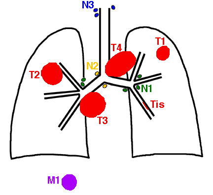
In the diagram below utilizing a lung carcinoma as an example, the principles of staging are illustrated:
| Staging of Malignant Neoplasms |
| Stage |
Definition |
| Tis |
In situ, non-invasive (confined to epithelium)
原位,无浸润(局限于上皮内) |
| T1 |
Small, minimally invasive within primary organ site
原发部位较小 |
| T2 |
Larger, more invasive within the primary organ site
原发部位较大 |
| T3 |
Larger and/or invasive beyond margins of primary organ site
更大和或浸润超过了原发器官的边缘
|
| T4 |
Very large and/or very invasive, spread to adjacent organs
非常大和(或)浸润到邻近器官 |
| N0 |
No lymph node involvement
没有淋巴结转移 |
| N1 |
Regional lymph node involvement
局限性淋巴结转移 |
| N2 |
Extensive regional lymph node involvement
广泛的淋巴结转移 |
| N3 |
More distant lymph node involvement
更多远处淋巴结转移 |
| M0 |
No distant metastases
无远处转移(血道转移) |
| M1 |
Distant metastases present
远处转移(血道转移) |
|
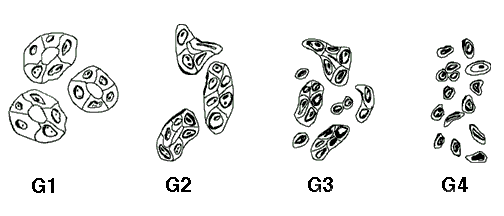 |
Grading(肿瘤分级)
Grading schema are based upon the microscopic appearance of a neoplasm with H&E staining. In general, a higher grade means that there is a lesser degree of differentiation and the worse the biologic behavior of a malignant neoplasm will be. A well-differentiated neoplasm is composed of cells that closely resemble the cell of origin, while poorly differentiated neoplasms have cells that are difficult to recognize as to their cell of origin. Grading schema have been devised for many types of neoplasms, mainly carcinomas. Most grading systems have three or four grades (designated with numbers or roman numerals).
In the diagram below utilizing an adenocarcinoma as an example, the principles of grading are illustrated:
| Grading of Malignant Neoplasms |
| Grade |
Definition |
| I |
Well differentiated (高分化) |
| II |
Moderately differentiated
中度分化 |
| III |
Poorly differentiated
低度分化 |
| IV |
Nearly anaplastic
未分化 |
肿瘤分级
肿瘤分级是对恶性肿瘤(主要是癌)而言的,通过显微镜观察HE染色切片可进行区别。一般来说,更高的分级说明肿瘤细胞分化较好,与起源组织更相似,生物学行为更好。常用的分级法为三级法或四级法。 |