
 |
Here is a small fibroadenoma of the breast. The blue dye was injected during a radiographic procedure to mark the location of the neoplasm so the surgeon could find it. 图示是乳腺纤维腺瘤。蓝染部位是放射引导下注射定位的瘤部位,便于手术。 |
 |
Remember that the most common neoplasm is a benign nevus (pigmented mole) of the skin, and most people have several, as seen here over the skin of the chest. As a general rule, benign neoplasms do not give rise to malignant neoplasms.
最常见的瘤是皮肤的黑色素痣,多数人都有。图示是胸部皮肤痣,这种良性瘤通常不会恶化。 |
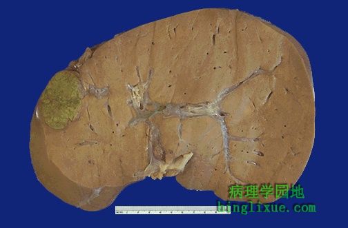 |
Here is a small hepatic adenoma, an uncommon benign neoplasm, but one that shows how well-demarcated an benign neoplasm is. It also illustrates how function of the normal tissue is maintained, because the adenoma is making bile pigment, giving it a green color.
图示一个小的良性肝腺瘤,一种不常见的良性瘤,可见周围界限清楚;也可以看到肝脏的功能得以维持,因为腺瘤被产生的胆汁染成绿色。 |
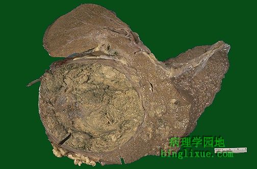 |
In contrast, this hepatocellular carcinoma is not as well circumscribed (note the infiltration of tumor off to the lower right) nor as uniform in consistency. It is also arising in a cirrhotic (nodular) liver. 相反,图示肝细胞癌既没有明显的界限,(已浸润到较远的右下方),也没有一致性。正发展成肝硬化。 |
 |
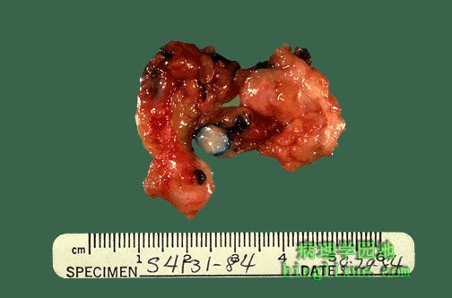
This renal cell carcinoma demonstrates distortion and displacement of the renal parenchyma by the tumor mass in the lower pole. This malignant neoplasm is variegated on cut surface, with yellow to white to red to brown areas. 图示:肾细胞癌 下面的巨大肿瘤块压迫肾实质,使其发生了扭曲变形。恶性肿瘤切面的颜色多样,可以是黄、白、红、棕之间的颜色。 |
 |
This excision of skin demonstrates a malignant melanoma, which is much larger and more irregular than a benign nevus. 图示:皮肤恶性黑色素瘤 与良性的黑色素痣相比更大、外形不规则。 |
 |
This is an example of metastases to the liver. Note that the tan-white masses are multiple and irregularly sized. A primary neoplasm is more likely to be a solitary mass. Metastasis is the best indication that a neoplasm is malignant. 图示:转移至肝脏的恶性肿瘤 可见大量灰白色大小不等的肿瘤结节,然而原发瘤多为单发。转移是恶性肿瘤的一个重要特征。 |
 |
This computed tomographic (CT) scan without contrast of the abdomen in transverse view demonstrates multiple mass lesions resulting in a markedly enlarged liver extending from right to nearly the left side of the upper abdomen. These are metastases from a colonic adenocarcinoma. A normal sized spleen is seen at the lower left. 图示:结肠腺癌肝转移CT影像 腹部横断面CT扫描图像显示多个肿块,致使肝脏体积明显增大,从右侧几乎延伸至左侧边缘。左侧(图的右下角)下部可见正常大小的脾脏。 |
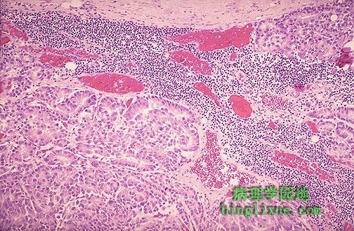 |
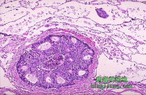
Microscopically, metastatic adenocarcinoma is seen in a lymph node here. It is common for carcinomas to metastasize to lymph nodes. The first nodes involved are those draining the site of the primary. 图示:腺癌淋巴结转移 癌通过淋巴道首先转移到初级淋巴结。 |
 |
Both lymphatic and hematogenous spread of malignant neoplasms is possible to distant sites. Here, a breast carcinoma has spread to a lymphatic in the lung. 图示:乳腺癌肺淋巴结转移 恶性肿瘤的淋巴道和血道转移可以发生在远隔器官。 |