
 |
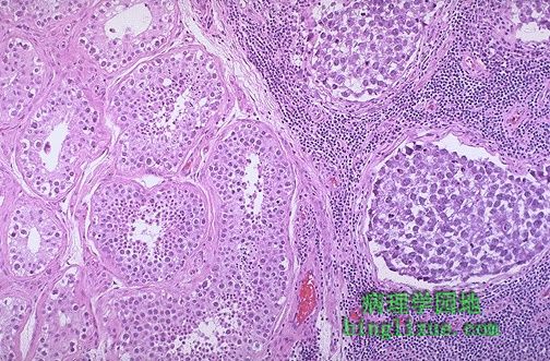
左侧为正常睾丸,右侧为精原细胞瘤。肿瘤细胞与正常的生殖细胞相比,大小和着色都不同。在精原细胞瘤癌巢中可见淋巴间质。 Normal testis appears at the left, and seminoma is present at the right. Note the difference in size and staining quality of the neoplastic nests of cells compared to normal germ cells. Note the lymphoid stroma between the nests of seminoma. |
 |
典型的精原细胞瘤组织学类型。肿瘤的细胞团间的间质以淋巴浸润为特征。精原细胞瘤癌细胞巨大、泡状核和胞浆疏松。 This is the histologic pattern of the typical seminoma. Lobules of neoplasitic cells have an intervening stroma with characteristic lymphoid infiltrates. The seminoma cells are large with vesicular nuclei, and pale watery cytoplasm. |
 |
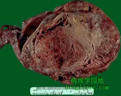
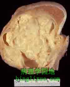
睾丸胚胎癌,上方是正常的睾丸的边缘。比精原细胞瘤质软,而且色彩更多样化,从红色到褐色,再到棕色,可见明显的出血和坏死。 Here is an embryonal carcinoma of the testis. There is a rim of normal testis superiorly. The tumor is soft and much more variegated than the seminoma, with red to tan to brown areas, including prominent hemorrhage and necrosis. |
 |
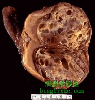
更大的睾丸肿瘤,主要由胚胎癌组成,但散在质硬的白色区域在组织学上是畸胎瘤。因此,这个睾丸肿瘤是胚胎癌和畸胎瘤的混合体(往往叫做恶性畸胎瘤)。胚胎癌比精原细胞瘤侵袭性更强。AFP(甲胎蛋白)通常升高。 Here is an even larger testicular neoplasm. It is composed mostly of embryonal carcinoma, but there are scattered firmer white areas that histologically are teratoma. Thus, this testicular neoplasm is mixed embryonal carcinoma plus teratoma (sometimes called teratocarcinoma). Embryonal carcinoma is more aggressive than seminoma. The alpha- fetoprotein is often elevated. |
 |
伴有畸胎瘤的胚胎癌,来自畸胎瘤成分的岛状的蓝白色软骨是清楚可见。左侧是正常的棕色睾丸的边缘。 Here is an embronal carcinoma mixed with teratoma in which islands of bluish white cartilage from the teratoma component are more prominent. A rim of normal brown testis appears at the left. |
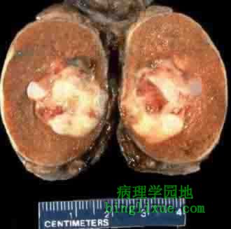 |
小睾丸癌,红白色的肿块组织伴有带蓝色的软骨。另外在显微镜下主要是畸胎瘤,但也有胚胎癌存在。 A small testicular carcinoma is shown here. There is a mixture of bluish cartilage with red and white tumor tissue. This neoplasm microscopically contained mainly teratoma, but areas of embryonal carcinoma were also present. |
 |
主要成分为畸胎瘤的睾丸肿瘤,但在显微镜下胚胎癌和精原细胞瘤都可找到。与卵巢相比,睾丸中单纯良性畸胎瘤是非常少见的。 Here is a testicular neoplasm that is mostly teratoma, but embryonal carcinoma and seminoma were found microscopically. In contrast with the ovary, pure benign teratomas of the testis are very rare. |
 |
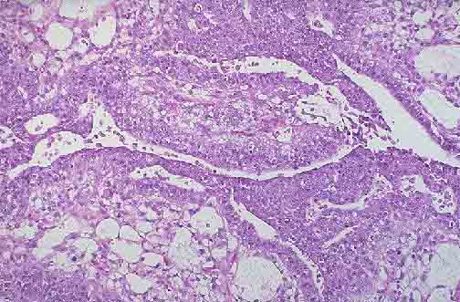
胚胎癌的组织学类型,一片蓝色的细胞正在形成原始的小管。 This is the histologic pattern of embryonal carcinoma. Sheets of blue cells are trying to form primitive tubules. |
 |
图下部是软骨,其上是由间叶细胞组成的原始间质,左侧是胚胎癌最有特点的原始细胞。这是混有畸胎瘤的胚胎癌。 At the bottom is a focus of cartilage. Above this is a primitive mesenchymal stroma and to the left a focus of primitive cells most characteristic for embryonal carcinoma. This is embryonal carcinoma mixed with teratoma. |
 |
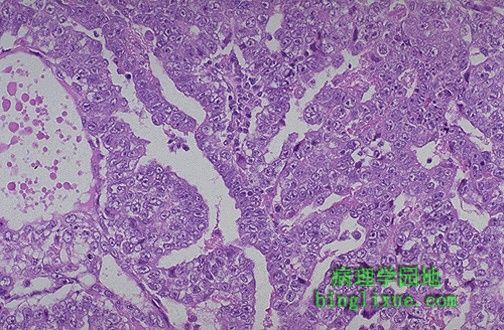
睾丸的内胚层窦肿瘤(卵黄囊肿瘤)由原始生殖细胞构成,这些细胞形成血管球或胚芽样结构。儿童多发,但总体来说是少见的。 An endodermal sinus tumor (yolk sac tumor) of the testis is shown composed of primitive germ cells that form glomeruloid or embryonal-like structures. These tumors are most frequent in children, but overall they are rare. |