
 |
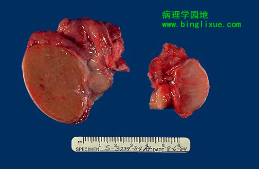
左图为正常睾丸,右图为萎缩睾丸。双侧睾丸萎缩在多种情况下都能发生,包括慢性酒精中毒、垂体功能减退、动脉粥样硬化、化疗或放疗以及严重的迁延性疾病。另外,隐睾、炎症也可导致萎缩。腮腺炎也可引起睾丸炎及其萎缩。 On the left is a normal testis. On the right is a testis that has undergone atrophy. Bilateral atrophy may occur with a variety of conditions including chronic alcoholism, hypopituitarism, atherosclerosis, chemotherapy or radiation, and severe prolonged illness. A cryptorchid testis will also be atrophic. Inflammation may lead to atrophy. Mumps, the most common cause for orchitis, usually has a patchy pattern of involvement that does not lead to sterility. |
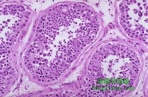 |
正常睾丸显微镜下结构,生精小管中有大量的生殖细胞,支持细胞是难以觉察的。小的暗黑色长形精子在管中央能见到。 This is the microscopic appearance of normal testis. The seminiferous tubules have numerous germ cells. Sertoli cells are inconspicuous. Small dark oblong spermatozoa are seen in the center of the tubules. |
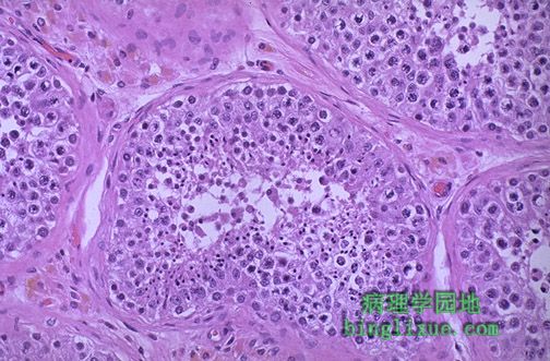 |
间质组织中可见粉红色的间质细胞。同时可见淡黄*色褐色素。那里正有精子形成。 Pink Leyding cells are seen here in the interstitium. Note the pale golden brown pigment as well. There is active spermatogenesis. |
 |
右上方可见生精小管的灶状萎缩。儿童感染腮腺炎病毒可致睾丸炎,是这种情况最常见的原因。不过这种感染通常不会引起睾丸明显萎缩影响精子的数量。 There is focal atrophy of tubules seen here to the upper right. The most common reason for this is probably childhood infection with the mumps virus, which produces a patchy orchitis. However, it is unusual for this infection to cause enough atrophy to significantly affect the sperm count. |
 |
萎缩的睾丸,可见生殖细胞显著减少而剩下高柱状粉红色的支持细胞,管周及间质纤维化。如果病变广泛将导致不育。想得到孩子但不育的夫妻中约有一半是因为男性生殖系统存在问题。 Atrophic testis is demonstrated here. Note the marked loss of germ cells with remaining tall pink Sertoli cells, peritubular fibrosis, and interstitial fibrosis. If generalized, this is a cause for infertility. About half the time when infertility occurs in couples wanting children, the cause is a problem in the male genital system. |
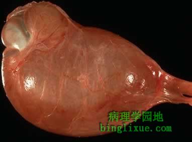 |
明显的鞘膜积液,此种病变相当常见。因各种各样的炎性和肿瘤的原因,透亮液体在由浆膜围绕形成的睾丸鞘膜囊中聚集。透视检测可区别积液与真正的睾丸组织,因积液能透光,而睾丸组织不能透光。 Here is a large hydrocele of the testis. Such hydroceles are fairly common. Clear fluid accumulates in a sac of tunica vaginalis lined by a serosa with a variety of inflammatory and neoplastic conditions. A hydrocele must be distinguished from a true testicular mass, and transillumination may help, because the hydrocele will transilluminate but a testicular mass will be opaque. |
 |
睾丸因扭转而梗死,扭转罕见,但是医疗紧急事件。精索扭转阻断静脉回流就会导致出血性梗死。下降不完全或阴囊韧带缺乏使睾丸活动度增加即可能更易发生扭转。尽快的外科手术,解开并在适当的地方缝合精索可防止以后再发生扭转,从而防止梗死。 This testis has undergone infarction following testicular torsion. Torsion is an uncommon condition, but a medical emergency. It occurs when twisting of the spermatic cord cuts off the venous drainage, leading to hemorrhagic infarction. Greater mobility from incomplete descent or lack of a scrotal ligament predisposes to this condition. Immediate treatment by surgically untwisting and suturing the cord in place to prevent future torsion will prevent infarction. |
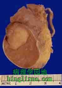 |
睾丸中见到肿块病变是精原细胞瘤。生殖细胞肿瘤是睾丸肿瘤中最常见的类型。在15~34年龄段中最常见。通常有以下几种组织学构成:精原细胞瘤、胚胎癌、畸胎瘤、绒毛膜癌。精原细胞瘤是最可能只有一种组织学类型的,如这个睾丸中所见。 The mass lesion seen here in the testis is a seminoma. Germ cell neoplasms are the most common types of testicular neoplasm. They are most common in the 15 to 34 age group. They often have several histologic components: seminoma, embryonal carcinoma, teratoma, choriocarcinoma. The one that is most likely to be of one histologic type is seminoma, as in the testis seen here. |
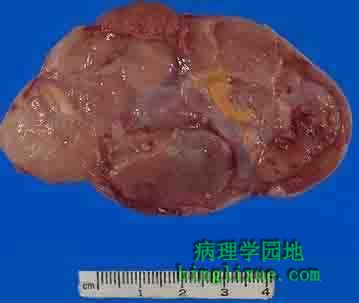 |
另例睾丸精原细胞瘤。最右边是残留正常睾丸的少量边缘。肿块质软、棕褐色、分叶状。 Here is another seminoma of the testis. A small rim of remaining normal testis appears at the far right. The tumor is composed of lobulated soft tan to brown tissue. |
 |
更大的精原细胞瘤,正常睾丸在肿块左侧,并且精索延伸到那里。因患者的恐惧和消极心理,没有尽早检查和治疗,因此肿瘤长的很大。 Here is a seminoma that is larger yet. Normal testis appears to the left of the mass, and the spermatic cord extends to the left of that. The size of this neoplasm demonstrates the factors of fear and denial that occur in many patients, delaying detection and therapy. |