
 |
根治性前列腺切除术所得的前列腺标本,展现的是后部的(上图可见)不规则黄*色小结节。证实为前列腺腺癌。含有腺癌的前列腺体不一定增大。另外,腺癌可与增生共存。不过,前列腺增生不是癌前病变。前列腺腺癌的分级是根据肿瘤的浸润程度而定的。 These sections through a prostate removed via radical prostatectomy reveal irregular yellowish nodules, mostly in the posterior portion (seen here superiorly). This proved to be prostatic adenocarcinoma. Prostate glands containing adenocarcinoma are not necessarily enlarged. Adenocarcinoma may also coexist with hyperplasia. However, prostatic hyperplasia is not a premalignant lesion. Staging of prostatic adenocarcinoma is based upon how extensive the tumor is. |
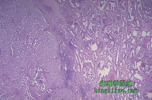 |
右侧是正常前列腺,可见散在分布的淀粉样小体;左侧是前列腺癌,可见癌腺体小拥挤。前列腺癌的组织学分级(如Gleason氏分级系统最常使用)。例如,此腺癌的Gleason分级为3/3。 At the right are normal prostatic glands containing scattered corpora amylacea. At the left is prostatic adenocarcinoma. Note how the glands of the carcinoma are small and crowded. Prostatic adenocarcinomas are given a histologic grade (Gleason's grading system is used most often, and includes a score of 1 to 5 for the most prominent component added to a score of 1 to 5 for the next most common pattern). For example, this adenocarcinoma could be given a Gleason of 3/3. |
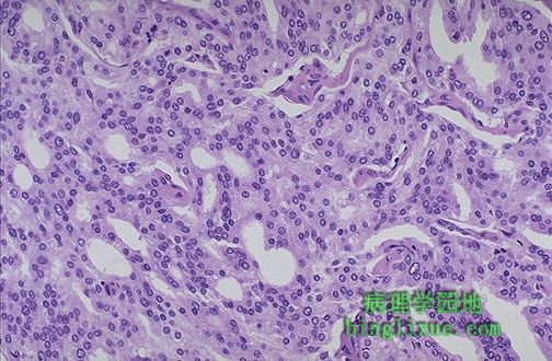 |
高倍镜,前列腺癌中腺体仍可辨认,但其间没有间质且核染色过深。 At high magnification, the neoplastic glands of prostatic adenocarcinoma are still recognizable as glands, but there is no intervening stroma and the nuclei are hyperchromatic. |
 |
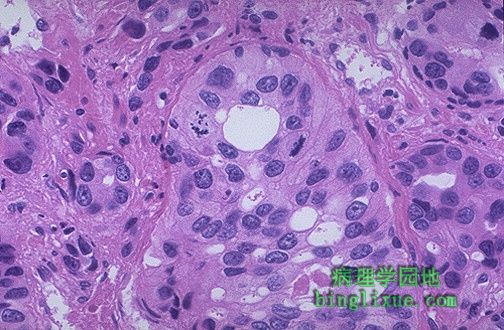
细胞核中可见明显核仁是前列腺癌典型的特征。 Prominent nucleoli are seen in the nuclei of this prostatic adenocarcinoma, which is a characteristic feature. |
 |
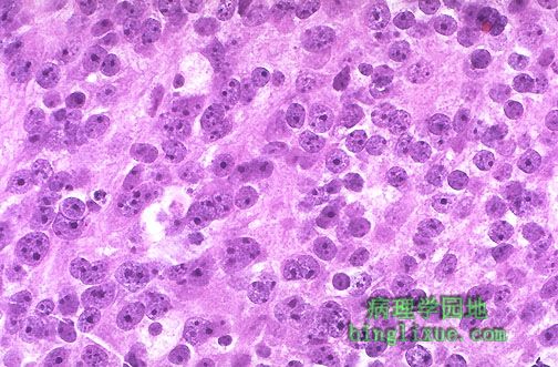
高倍镜下低分化的前列腺癌的细胞中可见核仁和核分裂像。 At high magnification, this poorly differentiated prostatic adenocarcinoma demonstrates cells with nucleoli and mitotic figures. |
|
前列腺癌分化程度极低,腺体结构不能分辨,仅有成行的细胞浸润。 This adenocarcinoma of prostate is so poorly differentiated that no glandular structure is recognizable, only cells infiltrating in rows. |
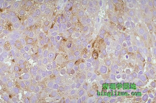 |
前列腺癌免疫过氧化物酶染色显示前列腺特异抗原(PSA)阳性。PSA因其用于对可能患有前列腺癌的男性作血清检测而闻名。 By immunoperoxidase staining with antibody to prostate specific antigen (PSA), this adenocarcinoma of prostate shows positivity. PSA is better known as a serum test to detect males that may have prostate cancer. |
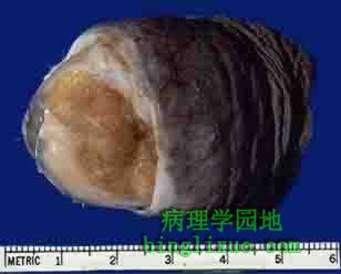 |
阴*茎龟头的鳞状细胞癌。可见未进行包皮切除术,增加了发生癌的可能性。暗红色的肿块表面有溃疡形成。 Here is a squamous cell carcinoma of the head of the penis. Note the uncircumcised state, which increases the risk for such carcinomas. The neoplasm is reddish-tan with an ulcerated surface. |
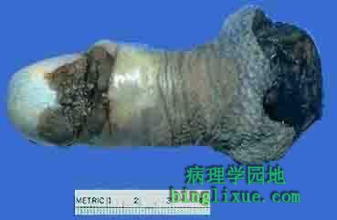 |
阴*茎切除术后的标本显示鳞状细胞癌为大红褐色霉菌状生长的团块。 This is a squamous cell carcinoma of the penis (penectomy specimen) that is a larger reddish brown fungating mass. |
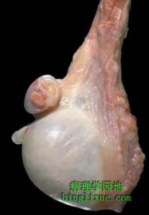 |
正常的睾丸及附属结构。可见睾丸、附睾和精索。可见2个退化的结构,睾丸附件和附睾附件。 Here is a normal testis and adjacent structures. Identify the body of the testis, epididymis, and spermatic cord. Note the presence of two vestigial structures, the appendix testis and the appendix epididymis. |