
 |
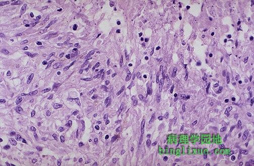
These are epithelioid cells around the center of a granuloma. They get their name from the fact that they have lots of pink cytoplasm similar to squamous epithelial cells. Their nuclei tend to be long and stringy. 图示上皮样细胞围绕在肉芽肿中央周围。由于这些细胞有丰富的粉红色胞浆类似于鳞状上皮细胞而得名。细胞核呈长梭形。 |
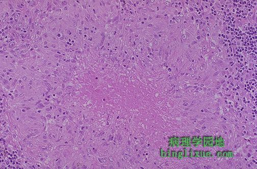 |
This is a caseating granuloma. Epithelioid cells surround a central area of necrosis that appears irregular, amorphous, and pink. Grossly, areas of caseation appear cheese-like. 图示干酪样肉芽肿。上皮细胞围绕在中央坏死区域周围,坏死物呈不规则、无定形、粉红色,干酪样坏死区状似奶酪。 |
 |
Granulomas caused by Mycobacterium tuberculosis and by pathogenic fungi such as Histoplasma capsulatum or Cryptococcus neoformans are often caseating. Here, the area of caseation is seen at the upper right. 图示:干酪样坏死(右上角) 肉芽肿可以由结核分枝杆菌和其它的病原菌如:荚膜组织胞浆菌或新型隐球菌。 |
 |
With a poor immune response to the agents producing granulomatous inflammation, there is extensive spread of infection with the production of a "miliary" pattern of granulomas as seen here in the lung of a patient with miliary tuberculosis. The 1 to 2 mm granulomas are scattered around like millet seeds. 图示:粟粒性结核病 免疫反应较差时,病变可呈粟粒样播散,如图可见肺组织中大量散在的直径1-2mm的粟粒样肉芽肿。 |
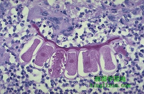 |
Here is a foreign body type giant cell at the upper left of center adjacent to a segment of vegetable material aspirated into the lung. Such foreign body giant cells have nuclei scattered haphazardly about the cell. 图示:异物巨细胞(左上方) 细胞核散在不规则分布。 |
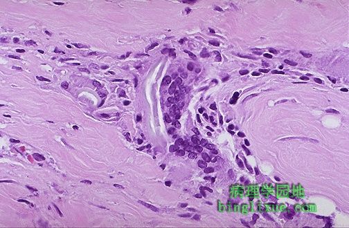 |
Two foreign body giant cells are seen just to the right of center where there is a bluish strand of suture material from a previous operation. 两个异物巨细胞位于中央偏右,同时可见以前手术留下的浅蓝色的缝线。 |
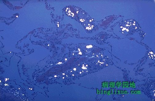 |
Seen under polarized light are numerous bright white crystals of talc in a patient who was an intravenous drug user. The injected drug was diluted with the talc. Such foreign material can produce a granulomatous reaction. 静脉给药病人偏振光显示亮白色滑石结晶。注入的药物用滑石稀释。这样的异物可促使肉芽肿形成。 |
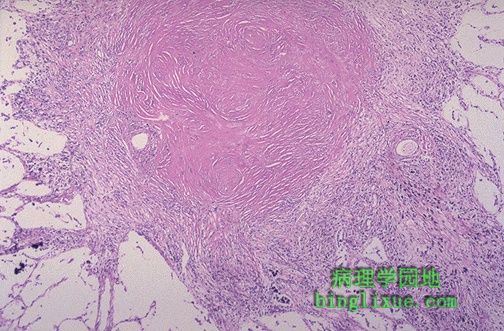 |
Sometimes the inflammatory reaction is mainly one of scarring, as seen here with a silicotic nodule of the lung. The inhaled silica persists indefinitely and produces an inflammatory reaction that is marked by prominent fibrosis. Dense pink collagen is seen in the center of the nodule. 图示:硅结节 长期吸入二氧化硅致肺纤维化,如图红节中央可见高密度粉红色胶原。 |