
 |
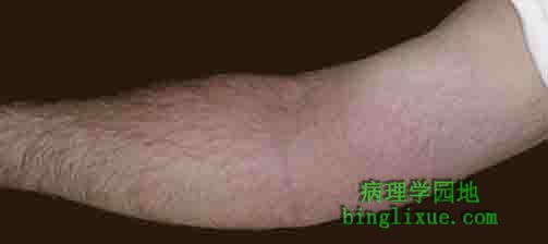
The cardinal signs of inflammation are rubor (redness), calor (heat), tumor (swelling), dolor (pain), and loss of function. Seen here is skin with erythema, compared to the more normal skin at the far right. 炎症的主要局部表现是红、肿、热、痛、功能障碍。图示:可见皮肤红斑,右侧为正常皮肤。 |
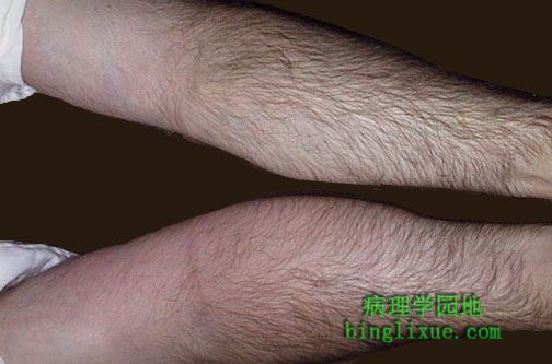 |
The arm at the bottom is swollen (edematous) and reddened (erythematous) compared to the arm at the top. 下面变红、肿胀,上面为正常。 |
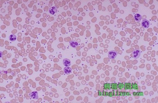 |
Acute inflammation is marked by an increase in inflammatory cells. Perhaps the simplest indicator of acute inflammation is an increase in the white blood cell count in the peripheal blood, here marked by an increase in segmented neutrophils (PMN's). 图示:外周血嗜中性粒细胞 急性炎症时炎细胞往往增多。在外周血中可以见到明显白细胞增多。 |
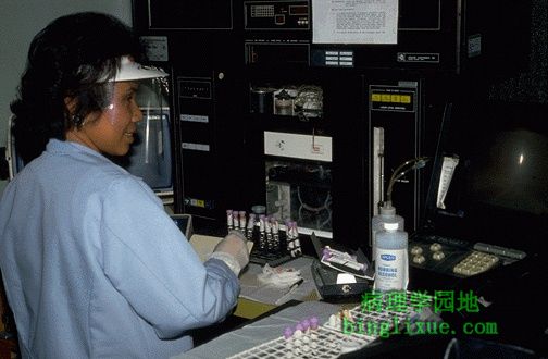 |
Here is a laboratory instrument called a Coulter Counter that enumerates the white blood cells, red blood cells, and platelets in the peripehral blood. A "CBC" (complete blood count) is a commonly ordered labortory test that is done on this instrument and gives important data, particularly in regard to WBC's. 图示:库尔特粒度仪。 用于计数白细胞、红细胞、血小板。整体血液指数是一项常常进行的检测的指标,可以在这样的仪器上完成,给出重要的数据,尤其是白细胞的计数。 |
 |
Seen here is vasodilation with exudation that has led to an outpouring of fluid with fibrin into the alveolar spaces, along with PMN's. The series of events in the process of inflammation are:
|
 |
As in the preceding diagram, here PMN's that are marginated along the dilated venule wall (arrow) are squeezing through the basement membrane (the process of diapedesis) and spilling out into extravascular space. 图示:嗜中性粒细胞经变形运动通过血管壁基膜并渗出到血管外。称为炎细胞渗出。 |
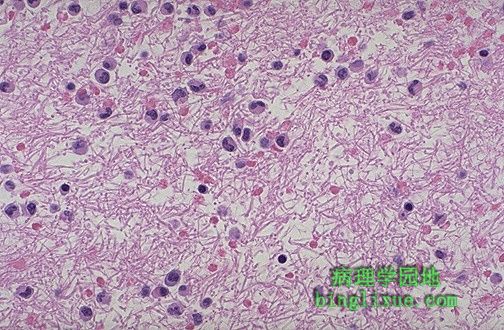 |
Here is an example of the fibrin mesh in fluid with PMN's that has formed in the area of acute inflammation. It is this fluid collection that produces the "tumor" or swelling aspect of acute inflammation. 图示:在急性炎症病灶,有嗜中性粒细胞的渗出液含有大量纤维蛋白,液体的聚集是导致局部肿胀的重要原因。 |
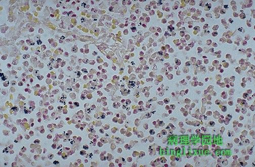 |
This tissue gram stain of an acute pneumonia demonstrates gram positive cocci that have been eaten by the numerous PMN's exuded into the alveolar space. Opsonins such as IgG and C3b facilitate the attachment of PMN's to offending agents such as bacteria so that the PMN's can phagocytose them. 图示:革兰氏染色显示大量渗出到肺泡腔的嗜中性粒细胞吞噬了革兰阳性菌。调理素(如IgG和补体C3b)能促进嗜中性粒细胞粘附破坏病原如细菌,从而使嗜中性粒细胞能有效的吞噬病原。 |
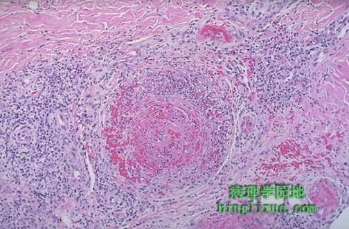 |
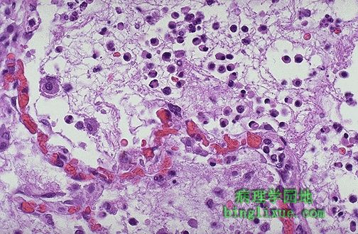
The vasculitis shown here demonstrates the destruction that can accompany the acute inflammatory process and the interplay with the coagulation mechanism. The arterial wall is undergoing necrosis, and there is thrombus formation in the lumen. 图示:脉管炎损伤性病变在于急性炎症和凝血机制的共同作用。动脉壁逐渐坏死,血管腔内有血栓形成。 |
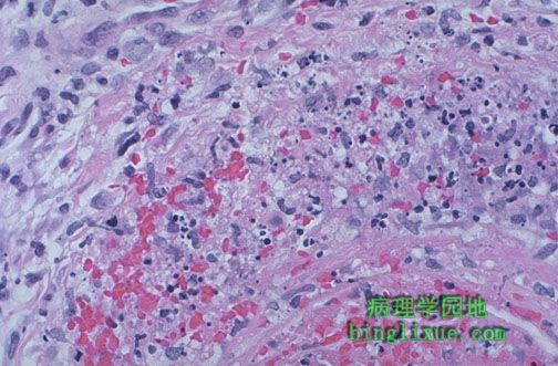 |
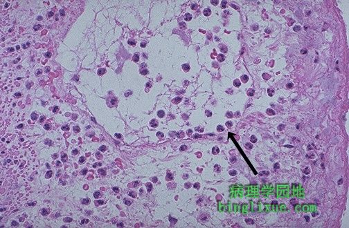
At higher magnification, vasculitis with arterial wall necrosis is seen. Note the fragmented remains of neutrophilic nuclei (karyorrhexis). Acute inflammation is a non-selective process that can lead to tissue destruction. 高倍镜下,脉管炎患者动脉壁坏死,可见嗜中性粒细胞核碎片(核碎裂)。急性炎症是一种非选择性的病理过程,可导致组织的损伤。 |