
 |
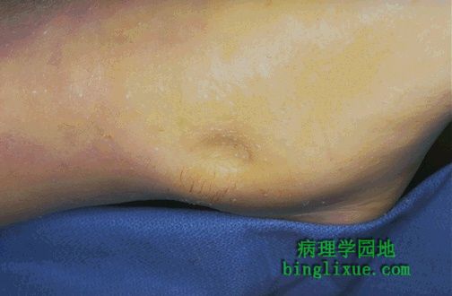
Here is simple edema, or fluid collection within tissues. This is "pitting" edema because, on physical examination, you can press your finger into the skin and soft tissue and leave a depression. 图示:凹陷性水肿 液体在组织间隙积聚形成水肿。体格检查时,手指按压相应皮肤和软组织,手指离开后凹陷不能立即恢复。 |
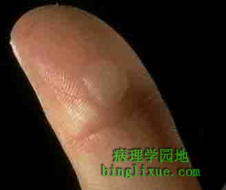 |
This example of a fluid collection, a friction blister of the skin, is an almost trivial example of edema. 皮肤过度摩擦,使皮下液体积聚,这样的水肿对机体的影响很小。 |
 |
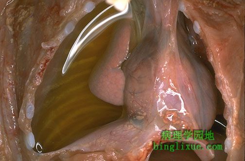
This example of edema with inflammation is not trivial at all: there is marked laryngeal edema such that the airway is narrowed. This is life-threatening. Thus, fluid collections can be serious depending upon their location. 图示:喉头水肿 炎症发生的喉头水肿对机体的影响较大,引起气道狭窄,甚至威胁生命。水肿对机体的影响与发生部位密切相关。 |
 |
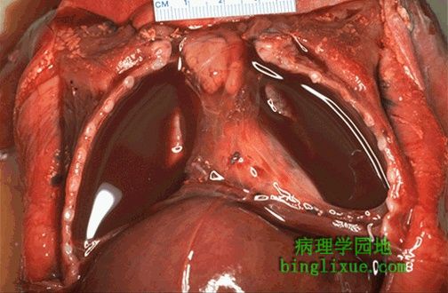
Here is an example of fluid collection into a body cavity, or an effusion. This is a right pleural effusion (in a baby). Note the clear, pale yellow appearance of the fluid. This is a serous effusion. Extravascular fluid collections can be classified as follows: 小儿胸腔积液,液体呈浅黄*色,渗出严重。血管外液体的积聚分为以下几种类型: Exudate: extravascular fluid collection that is rich in protein and/or cells. Fluid appears grossly cloudy. Transudate: extravascular fluid collection that is basically an ultrafiltrate of plasma with little protein and few or no cells. Fluid appears grossly clear. Effusions into body cavities can be further described as follows:
|
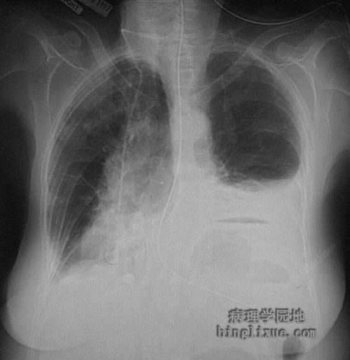 |
This radiograph demonstrates fluid in the left pleural cavity. This pleural effusion could result from a transudate (serous effusion) or from hemorrhage (hemothorax), or serous fluid tinged with blood (serosanguinous effusion). This effusion could be chylous (which is quite rare). A purulent exudate at this location may be termed empyema. An air-fluid level is seen in the stomach below the dome of the left diaphragmatic leaf. 图示:X线示左胸膜腔积液 |
 |
Here is an example of bilateral pleural effusions. Note that the fluid appears reddish, because there has been hemorrhage into the effusion. This is a serosanguinous effusion. 图示:双侧胸膜腔积液 可见红色血性液体,此为血性浆液渗出的结果。 |
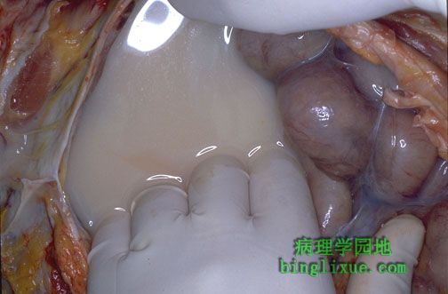 |
The milky white fluid shown here in the peritoneal cavity represents a chylous ascites. This is an uncommon fluid accumulation that can be due to blockage of lymphatic drainage, in this case by a malignant lymphoma involving the mesentery and retroperitoneum. 图示:腹膜腔乳糜性腹水,呈乳白色。这是一种特殊的液体积聚,发生于淋巴管阻塞,通常是恶性淋巴瘤累及肠系膜和腹膜后腔。 |
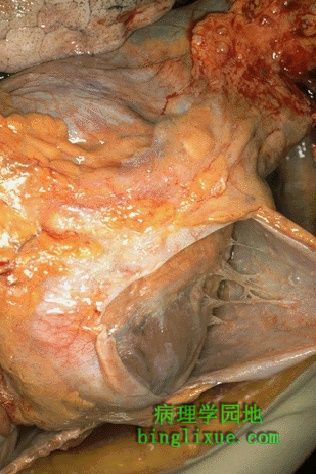 |
Exudation of a protein-rich fluid into a cavity leads to a transudate. The fibrin in this fluid can form a fibrinous exudate on the surfaces. Here, the pericardial cavity has been opened to reveal a fibrinous pericarditis with strands of stringy pale fibrin between visceral and parietal pericardium. 渗出液中蛋白质含量较高,会导致体腔积液。如果以纤维素为主,会导致粘膜表面的纤维素性炎症。图示纤维素性心包炎,在心包脏层和壁层之间可见白色绒毛状的纤维素。 |
 |
Microscopically, the fibrinous exudate is seen to consist of pink strands of fibrin jutting from the pericardial surface at the upper left. Below this, there are a few scattered inflammatory cells. 镜下可在左上方可见渗出的粉红色束状纤维突出于心包膜表面。其下面可见散在的炎细胞。 |
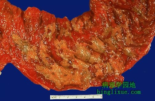 |
This yellow-green exudate on the surface of an inflamed, hyperemic (erythematous) bowel mucosa consists of many neutrophils along with fibrin and amorphous debris from dying cells. 炎性充血的肠粘膜表面可见黄绿色渗出物,由在病灶处纤维和坏死细胞碎片周围有许多中性粒细胞组成。 |