
 |
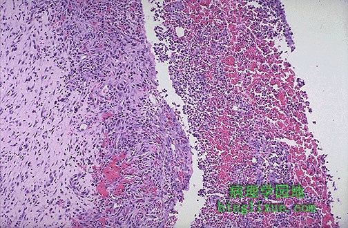
The wall of an abscess that is organizing has granulation tissue, seen here at the left. The purulent exudate with some hemorrhage is seen at the right in the abscess center. 图左侧可见正在机化的脓肿壁中有肉芽组织。右侧脓肿中央可见伴出血的脓性分泌物渗出。 |
 |
At high magnification, granulation tissue has capillaries, fibroblasts, and a variable amount of inflammatory cells (mostly mononuclear, but with the possibility of some PMN's being present). 高倍速镜:肉芽组织由毛细血管、成纤维细胞和大量的各种炎细胞组成。炎细胞主要是单核细胞,也可以伴有嗜中性粒细胞的出现。 |
 |
The end result of inflammation can be scarring. Here, the alveolar walls are thickened and filled with pink collagen following an autoimmune disease lasting for decades. 炎症的结局是形成瘢痕。图示:几十年的自身免疫性疾病使得肺泡壁增厚、粉红色胶原纤维增生。 |
 |
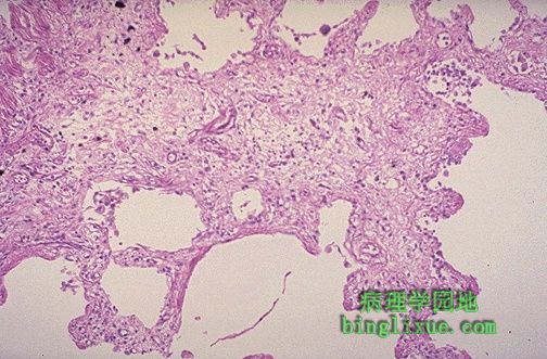
This is a healing biopsy site on the skin seen a week following the excision, The skin surface has re-epithelialized, and below this is granulation tissue with small capillaries and fibroblasts forming collagen. After a month, just a small collagenous scar will remain. 局部切除一周后皮肤愈合部位活检显示:皮肤表面已经有再生的上皮,其下部为含有毛细血管和成纤维细胞的肉芽组织,一个月后转变为胶原纤维瘢痕。 |
 |
Granulomatous disease can become quite extensive. Here are numerous confluent granulomas in a case of pulmonary tuberculosis. 肉芽肿性疾病是十分常见的。图示:肺结核病大量融合性肉芽肿。 |
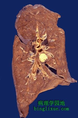 |
Grossly, a granuloma tends to be a focal lesion. Seen here in a hilar lymph node is a granuloma. Granulomas due to infection are often "caseating" because they have prominent caseous necrosis. 肉芽肿是较局限化的。图示:肺门淋巴结处可见肉芽肿。感染引起的肉芽肿通常称为干酪样化,是因为通常有典型的干酪样坏死。 |
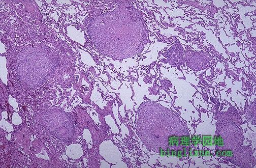 |
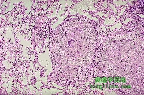
The focal nature of granulomatous inflammation is demonstrated in this microscopic section of lung in which there are scattered granulomas in the parenchyma. This is why the chest radiograph with tuberculosis or other granulomatous diseases is often described as "reticulonodular". A biopsy could miss such lesions from sampling error, too. 肺实质中可见数个散在的肉芽肿,由此可见肉芽肿的局限性特点。这也是肺结核病或其它肉芽肿疾病胸部X线检查被描述为网织结节状的原因。活检时采样错误也有可能导致检查不到这样的病变。 |
 |
Here are two pulmonary granulomas. Granulomatous inflammation typically consists of epithelioid macrophages, giant cells, lymphocytes, plasma cells, and fibroblasts. There may be some neutrophils. 图示:两个肺部肉芽肿。典型的肉芽肿由上皮样细胞、巨细胞、淋巴细胞、浆细胞和成纤维细胞组成,也可以有一定量的中性粒细胞。 |
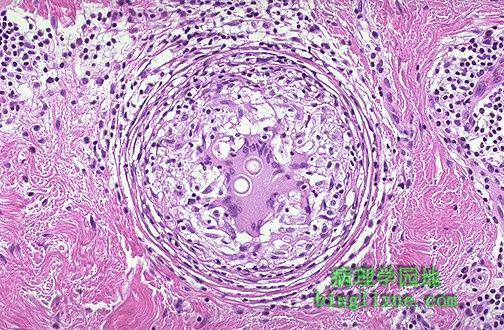 |
Granulomatous inflammation occurs in response to some agents which persist for a long time and require a more orchestrated immune response to fight them. The granuloma seen here demonstrates the typical rounded and focal nature of this type of inflammation. A couple of spherules of C. immitis are present in the giant cell in the center. 肉芽肿性炎常发生于病因长期作用于机体,同时较强的免疫反应也贯穿始终。图示典型的肉芽肿呈圆形局限性的特点。中央巨细胞中可见两个小球体。 |
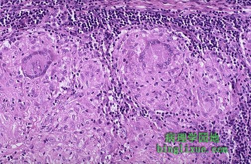 |
Giant cells are a "committee" of epithelioid macrophages. Seen here are two Langhans type giant cells in which the nuclei are lined up around the periphery of the cell. Additional pink epithelioid macrophages compose most of the rest of the granuloma. 巨细胞由多个上皮样细胞融合而成。图示两个朗格汉斯巨细胞,其细胞核沿着细胞周围排列。更多的上皮样细胞构成了肉芽肿的其它部分。 |