
 |
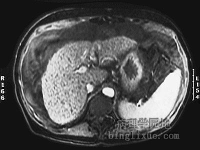
腹部横断面MRI影像显示体积小而有结节的硬化性肝脏。可见由门静脉高压导致的体积增大的脾脏。 This magnetic resonance imaging (MRI) scan of the abdomen in transverse view demonstrates a small, nodular liver with cirrhosis. The spleen is enlarged from portal hypertension |
 |
另例小结节型肝硬化。注意肝呈淡黄*色,表示脂肪变性(也由酒精引起)。 Here is another example of micronodular cirrhosis. Note that the liver also has a yellowish hue, indicating that fatty change (also caused by alcoholism) is present. |
 |
增强CT显示肝脏硬化缩小,脾增大(门静脉高压所致)。 This computed tomographic (CT) scan with contrast of the abdomen in transverse view demonstrates a small liver with cirrhosis. The spleen is enlarged from portal hypertension. |
 |
近观伴脂肪变的小结节型肝硬化可见小而呈黄*色结节。小结节型肝硬化也可见于Wilson病、原发性胆汁硬化和血色素沉着症。 A close-up view of a micronodular cirrhosis in a liver with fatty change demonstrates the small, yellow nodules. Micronodular cirrhosis may also be seen with Wilson's disease, primary biliary cirrhosis, and hemochromatosis. |
 |
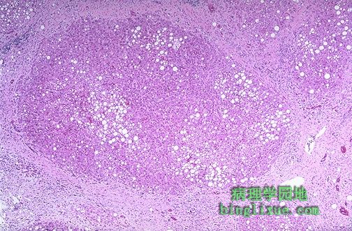
用显微镜观察肝硬化,肝细胞再生结节被桥接汇管区的纤维结缔组织包绕。结缔组织中散在淋巴细胞也有小胆管的增生。 Microscopically with cirrhosis, the regenerative nodules of hepatocytes are surrounded by fibrous connective tissue that bridges between portal tracts. Within this collagenous tissue are scattered lymphocytes as well as a proliferation of bile ducts. |
 |
伴中度脂肪变性的小结节型肝硬化。注意再生的肝细胞小结节被汇管区之间的纤维结缔组织包围。 Micronodular cirrhosis is seen along with moderate fatty change. Note the regenerative nodule surrounded by fibrous connective tissue extending between portal regions. |
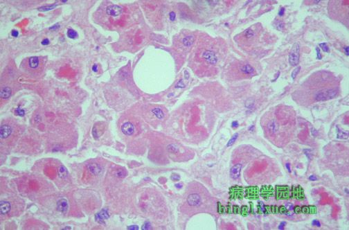 |
在高倍镜下能看到肝细胞内红色球形玻璃样物质,这是Mallory 玻璃样变,也称“酒精”玻璃样变,因为经常在慢性酒精性肝炎中能观察到。球形物质是肝细胞损伤时细胞浆中的中间丝聚集而成。 At high magnification can be seen globular red hyaline material within hepatocytes. This is Mallory's hyaline, also known as "alcoholic" hyaline because it is most often seen in conjunction with chronic alcoholism. The globules are aggregates of intermediate filaments in the cytoplasm resulting from hepatocyte injury. |
 |
此处能看到Mallory玻璃样变,但也有中性白细胞、肝细胞坏死、胶原沉积、脂肪变。这是典型的急性酒精性肝炎,发生于极度嗜酒的人(如短时间内大量饮酒)。 Mallory's hyaline is seen here, but there are also neutrophils, necrosis of hepatocytes, collagen deposition, and fatty change. These findings are typical for acute alcoholic hepatitis. Such inflammation can occur in a person with a history of alcoholism who goes on a drinking "binge" and consumes large quantities of alcohol over a short time. |
 |
肝硬化异常血流导致门脉高压。升高的压力传送至侧支静脉,侧支发生扩张。如图所示,肝硬化患者腹部扩张的静脉侧支形成了“海蛇头”样表现。 Portal hypertension results from the abnormal blood flow pattern in liver created by cirrhosis. The increased pressure is transmitted to collateral venous channels. Sometimes these venous collaterals are dilated. Seen here is "caput medusae" which consists of dilated veins seen on the abdomen of a patient with cirrhosis of the liver. |
 |
门脉高压使食管粘膜下静脉扩张时,会出现更严重的后果,称为食管静脉曲张。图示食管下段静脉曲张,即所示蓝色线状扩张的静脉。可见一处发生了出血。这些静脉容易被破坏,导致胃肠道大出血。 A much more serious problem produced by portal hypertension results when submucosal veins in the esophagus become dilated. These are known as esophageal varices. Varices are seen here in the lower esophagus as linear blue dilated veins. There is hemorrhage around one of them. Such varices are easily eroded, leading to massive gastrointestinal hemorrhage. |