
 |
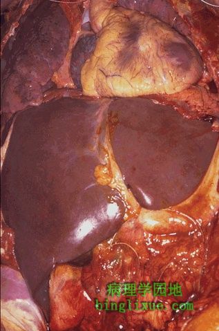
胸腔和腹腔的内脏图可见肝脏是最大的实质器官,位于膈下。右叶(图右侧)比左叶大。镰状韧带将两叶分开。 This is an in-situ photograph of the chest and abdominal contents. As can be seen, the liver is the largest parenchymal organ, lying just below the diaphragm. The right lobe (at the left in the photograph) is larger than the left lobe. The falciform ligament is the rough dividing line between the two lobes. |
 |
正常肝脏的外表面,褐色,表面光滑。正常的肝脏重约1200至1600克。 This is the external surface of a normal liver. The color is brown and the surface is smooth. A normal liver is about 1200 to 1600 grams. |
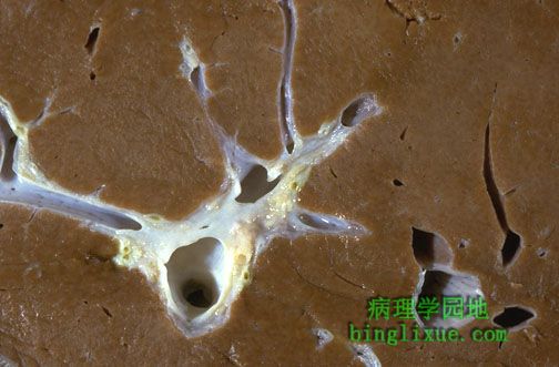 |
正常肝脏切面呈褐色,注意靠近门部,门静脉血液入肝脏,图中央左侧的门静脉分支,与肝动脉和胆管伴行。图右下方有一个肝静脉分支将肝脏的血液回流到下腔静脉。 The cut surface of a normal liver has a brown color. Near the hilum here, note the portal vein carrying blood to the liver, which branches at center left, with accompanying hepatic artery and bile ducts. At the lower right is a branch of hepatic vein draining blood from the liver to the inferior vena cava. |
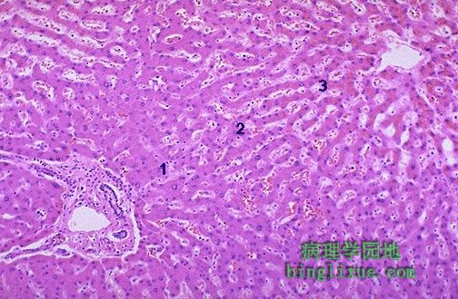 |
肝在组织学上分为无数的肝小叶。肝小叶中央是中央静脉(右上角),周围是汇管区(门管区)(左下角)。在功能上,肝脏可以从供氧的角度划分为3个区域。1区位于汇管区周围,含氧量较高,3区位于中央静脉周围,含氧量低,2区位于两者之间,含氧量一般。 Liver is divided histologically into lobules. The center of the lobule is the central vein. At the periphery of the lobule are portal triads. Functionally, the liver can be divided into three zones, based upon oxygen supply. Zone 1 encircles the portal tracts where the oxygenated blood from hepatic arteries enters. Zone 3 is located around central veins, where oxygenation is poor. Zone 2 is located in between. |
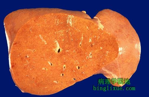 |
肝轻度肿大,被膜和切面呈黄白色,与肝脂肪变性一致。 This liver is slightly enlarged and has a pale yellow appearance, seen both on the capsule and cut surface. This uniform change is consistent with fatty metamorphosis (fatty change). |
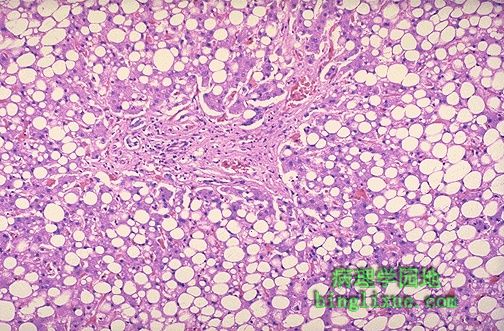 |
肝脂肪变的组织学表现。脂质在肝细胞内聚集,HE染色呈非常明显的空泡状。发达国家酒精中毒最重要的原因,而发展中国家重度营养不良是儿儿童的重要病因,另外,糖尿病、肥胖、严重的胃肠吸收障碍也是肝脂肪变的病因。 This is the histologic appearance of hepatic fatty change. The lipid accumulates in the hepatocytes as vacuoles. These vacuoles have a clear appearance with H&E staining. The most common cause of fatty change in developed nations is alcoholism. In developing nations, kwashiorkor in children is another cause. Diabetes mellitus, obesity, and severe gastrointestinal malabsorption are additional causes. |
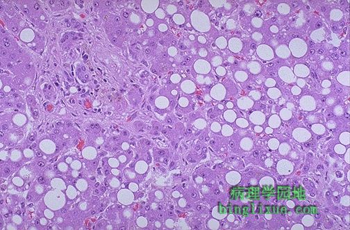 |
肝细胞的脂质空泡。当脂蛋白转运中断或脂肪酸堆积时,脂质就会聚集起来。乙醇是最常见的病因,作为肝毒素能阻碍肝细胞内的线粒体和微粒体的功能,导致脂质沉集。 Here are seen the lipid vacuoles within hepatocytes. The lipid accumulates when lipoprotein transport is disrupted and/or when fatty acids accumulate. Alcohol, the most common cause, is a hepatotoxin that interferes with mitochondrial and microsomal function in hepatocytes, leading to an accumulation of lipid. |
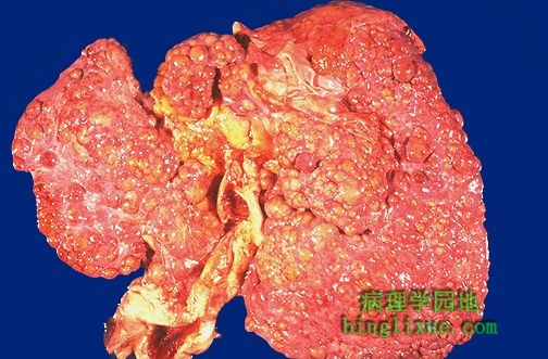 |
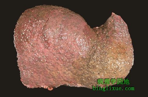
肝硬化是肝细胞坏死后,肝纤维化和肝细胞再生所致。产生结节,使肝脏变硬。图示结节大于3 mm,属大结节型肝硬化。 Ongoing liver damage with liver cell necrosis followed by fibrosis and hepatocyte regeneration results in cirrhosis. This produces a nodular, firm liver. The nodules seen here are larger than 3 mm and, hence, this is an example of "macronodular" cirrhosis. |
 |
另例大结节型肝硬化。病毒性肝炎(乙型或丙型)是大结节型肝硬化的最常见病因,Wilson’s病和alpha-1-抗胰蛋白酶缺陷也能产生肝大结硬化。 Here is another example of macronodular cirrhosis. Viral hepatitis (B or C) is the most common cause for macronodular cirrhosis. Wilson's disease and alpha-1-antitrypsin deficiency also can produce a macronodular cirrhosis. |
 |
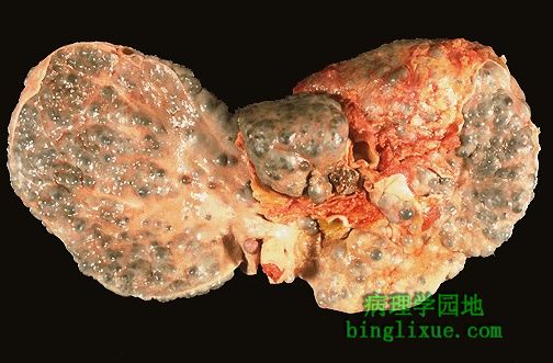
小结节型肝硬化,再生肝细胞结节平均小于3 mm,最常见的原因是慢性酒精性中毒,这种硬化需要数年。 This is an example of a micronodular cirrhosis. The regenerative nodules are quite small, averaging less than 3 mm in size. The most common cause for this is chronic alcoholism. The process of cirrhosis develops over many years. |