
 |
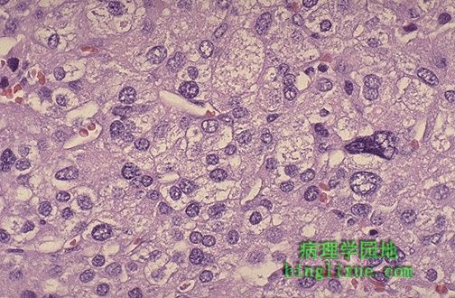
高倍镜下的肾上腺皮质癌,显示细胞的多形性、细胞核染色过深。无论是良性内分泌肿瘤还是恶性内分泌肿瘤都显示一定程度的细胞异型性,因此仅从组织学上很难区分肿瘤的良恶性。肿瘤越大,恶性的可能越大,但最好的诊断依据是肿瘤的浸润和转移。 Here is an adrenal cortical carcinoma seen microscopically at high power to demonstrate cellular pleomorphism with nuclear hyperchromatism. Both benign and malignant endocrine neoplasms demonstrate some degree of cellular pleomorphism, so it is not easy to tell benign from malignant on histologic grounds alone. The larger the neoplasm, the more likely it is malignant, but the best indicators are invasion and metastasis. |
 |
大的肾上腺肿瘤被切为两半,可见肿瘤呈现灰黑色,周围是被拉伸的黄*色皮质,右下为残余肾上腺。病人患有阵发性高血压。这是源于肾上腺髓质的肿瘤--嗜铬细胞瘤。 This large adrenal neoplasm has been sectioned in half. Note the grey-tan color of the tumor compared to the yellow cortex stretched around it and a small remnant of remaining adrenal at the lower right. This patient had episodic hypertension. This is a tumor arising in the adrenal medulla--a pheochromocytoma. |
 |
嗜铬细胞瘤显示嗜铬反应阳性,该肾上腺髓质肿瘤含有儿茶酚胺(肾上腺素和去甲肾上腺素)。下图是已被重铬酸盐固定的肿瘤标本,由于儿茶酚胺被氧化呈棕色。上图是未使用重铬酸盐固定的标本,肿瘤呈现粉红色到黄*色之间的颜色。 This pheochromocytoma demonstrates the chromaffin reaction. This neoplasm of the adrenal medulla contains catecholamines (epinephrine and norepinephrine). The section of tumor at the bottom has been placed into a dichromate fixative which turns the tissue brown as the catecholamines are oxidized. Compare to the section of pink to yellow tumor at the top which has not been placed in dichromate fixative. |
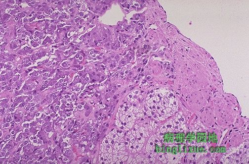 |
右下可见残存的肾上腺皮质,左上为颜色较暗的嗜铬细胞瘤细胞。 There is some residual adrenal cortical tissue at the lower center right, with the darker cells of pheochromocytoma seen above and to the left. |
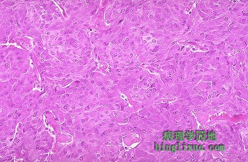 |
显微镜下,嗜铬细胞瘤由一些粉红到紫红的大细胞组成,在毛细血管间呈巢状分布。一想到嗜铬细胞瘤一定要记得10%这个数值:10%的肿瘤是双侧的,10%发生于儿童,10%的肿瘤是恶性的。 Microscopically, a pheochromocytoma is composed of large cells that are pink to mauve and arranged in nests with capillaries in between. Remember 10% when you think of a pheochromocytoma: 10% are bilateral, 10% are in children, 10% are malignant. |
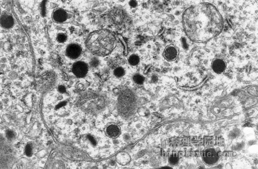 |
电子显微镜下,嗜铬细胞瘤的瘤细胞内包含神经内分泌颗粒,颗粒内含儿茶酚胺,胞质内所见到的黑色圆形小体即为此种颗粒。细胞核位于左上面。 By electron microscopy, the neoplastic cells of the pheochromocytoma contain neurosecretory granules. It is these granules that contain the catecholamines. The granules seen here appear as small black round objects in the cytoplasm of the cell. The cell nucleus is at the upper left. |
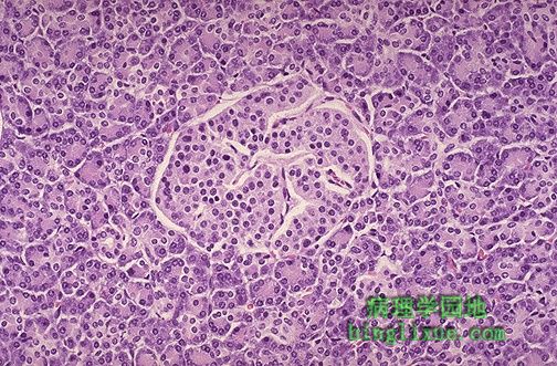 |
可见一正常胰岛被外分泌腺胰腺的腺泡包绕。胰岛包含分泌胰高血糖素的α(A)细胞 ,分泌胰岛素的β(B)细胞和分泌生长抑素的δ(D)细胞。 Here is a normal pancreatic islet of Langerhans surrounded by normal exocrine pancreatic acinar tissue. The islets contain alpha cells secreting glucagon, beta cells secreting insulin, and delta cells secreting somatostatin. |
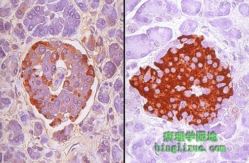 |
免疫过氧化物酶染色可帮助了解胰岛内细胞的特性。右图用胰岛素抗体识别α细胞。在图用胰高血糖素抗体识别β细胞。 Immunoperoxidase staining can help identify the nature of the cells present in the islets of Langerhans. On the right, antibody to insulin has been employed to identify the beta cells. On the left, antibody to glucagon identifies the alpha cells. |
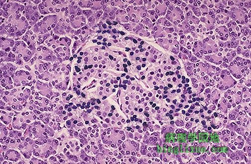 |
胰岛炎胰岛,病人最终将发展为I型糖尿病。水肿的胰岛内出现淋巴细胞浸润暗示自身免疫作用。胰岛的破坏导致胰岛素绝对缺乏,这是I型糖尿病的特征。 This is an insulitis of an islet of Langerhans in a patient who will eventually develop type I diabetes mellitus. The presence of the lymphocytic infiltrates in this edematous islet suggests an autoimmune mechanism for this process. The destruction of the islets leads to an absolute lack of insulin that characterizes type I diabetes mellitus. |
 |
胰岛出现粉红色的淀粉样变性,多见于II型糖尿病。 This islet of Langerhans demonstrates pink hyalinization (with deposition of amyloid) in many of the islet cells. This change is common in the islets of patients with type II diabetes mellitus. |