
 |
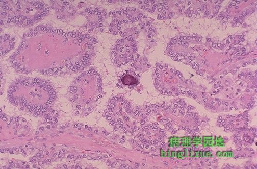
另例甲状腺乳头状腺癌。中央可见小砂粒体。肿瘤细胞的细胞核清晰可见。乳头状癌表现为无痛性肿块,存活时间长,甚至在转移的情况下也是如此。甲状腺乳头状癌转移最常见部位是颈部的局部淋巴结。事实上,一些乳头状癌可能首先发现的是结节状转移灶。 This is another papillary carcinoma of thyroid. Note the small psammoma body in the center. The cells of the neoplasm have clear nuclei. Papillary carcinomas are indolent tumors that have a long survival, even with metastases. The most favorite site of metastasis is to local lymph nodes in the neck. In fact, some papillary carcinomas may first present as nodal metastases. |
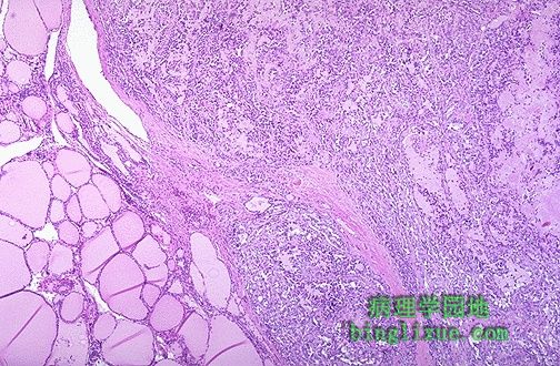 |
中间到右侧见甲状腺髓样癌。最右边呈现粉红色透明样,为淀粉样变性。此肿瘤来源于甲状腺C细胞,因此具有神经内分泌方面的特征,属APUD瘤,例如分泌降钙素。 At the center and to the right is a medullary carcinoma of thyroid. At the far right is pink hyaline material with the appearance of amyloid. These neoplasms are derived from the thyroid "C" cells and, therefore, have neuroendocrine features such as secretion of calcitonin. |
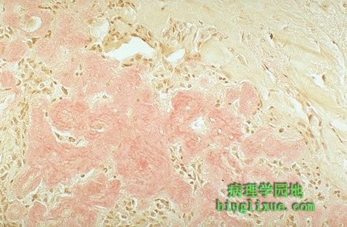 |
刚果红染色显示甲状腺髓样癌的淀粉样基质。髓样癌可以是散在单发,也可以是家族性发病。家族发病类型与多发性内分泌瘤综合征有关系。 Here the amyloid stroma of the medullary thyroid carcinoma has been stained with Congo red. Medullary carcinomas can be sporadic or familial. The familial kind are associated with multiple endocrine neoplasia syndrome. |
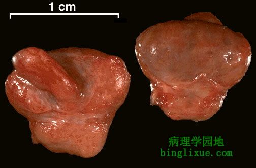 |
从蝶鞍切除的正常垂体。大部分是垂体前叶(腺垂体),朝向上方。左边图像显示垂体的上面观,下丘脑发出一蒂进入垂体。右边为垂体的下面观,垂体后叶(神经垂体)是位于底部较小的部分。 The normal gross appearance of the pituitary gland removed from the sella turcica is shown here. The larger portion, the anterior pituitary (adenohypophysis), is toward the top. The image at the left shows the superior aspect of the pituitary with the stalk coming from the hypothalamus entering it. The inferior aspect of the pituitary is shown at the right. The posterior pituitary (neurohypophysis) is the smaller portion at the bottom. |
 |
此处所示为正常垂体显微镜图像。腺垂体在右边,神经垂体在左边。 The normal microscopic appearance of the pituitary gland is shown here. The adenohypophysis is at the right and the neurohypophysis is at the left. |
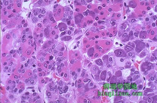 |
正常腺垂体显微镜图像。腺垂体包含嗜酸性细胞、嗜碱性细胞和嫌色细胞。染色方法多样,如要准确识别特定激素分泌物,免疫组织化学染色法是必要的。 The normal microscopic appearance of the adenohypophysis is shown here. The adenohypophysis contains three major cell types: acidophils, basophils, and chromophobes. The staining is variable, and to properly identify specific hormone secretion, immunohistochemical staining is necessary. A simplistic classification is as follows: |
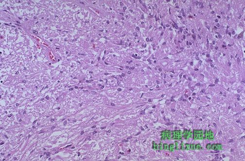 |
神经垂体类似神经组织,有神经胶质细胞、神经纤维、神经末梢及轴突内的神经内分泌颗粒。下丘脑(视上核和室旁核)形成的血管加压素(抗利尿激素,ADH)和催产素以神经内分泌颗粒的形式经轴突运送至神经垂体得以释放。 The neurohypophysis shown here resembles neural tissue, with glial cells, nerve fibers, nerve endings, and intra-axonal neurosecretory granules. The hormones vasopressin (antidiuretic hormone, or ADH) and oxytocin made in the hypothalamus (supraoptic and paraventricular nuclei) are transported into the intra-axonal neurosecretory granules where they are released. |
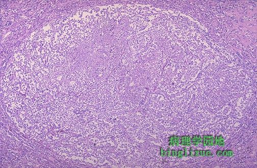 |
图示腺垂体微腺瘤,成年人发生机率为1~5%,很少出现明显的激素分泌导致临床症状和体征。 This is a microadenoma of the anterior pituitary. Such microadenomas may appear in 1 to 5% of adults. These microadenomas rarely have a significant hormonal output that leads to clinical disease. |
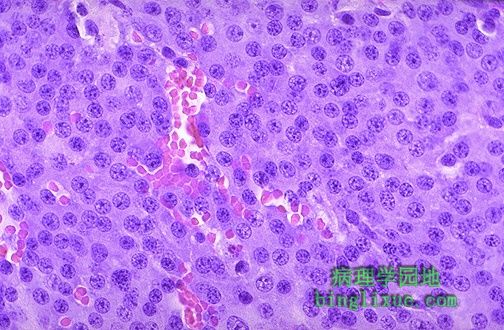 |
腺垂体腺瘤高倍镜图像, 内分泌肿瘤由一些圆形小细胞组成,细胞核小而圆,细胞质显示为粉红色到蓝色之间的颜色。细胞可排列成巢状或条索状,同时血管分布也较为丰富。 Here is a high power microscopic view of an adenohypophyseal adenoma. Endocrine neoplasms are composed of small round cells with small round nuclei and pink to blue cytoplasm. The cells may be arranged in nests or cords and endocrine tumors also have prominent vascularity. |
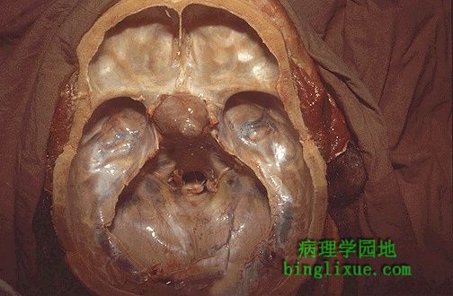 |
蝶鞍内的边界清楚的肿块为垂体腺瘤。尽管垂体腺瘤是良性的,但也能引起肿块压迫的后果:因压迫视交叉引起视力障碍、、头痛、产生催乳素ACTH等。 The circumscribed mass lesion present here in the sella turcica is a pituitary adenoma. Though pituitary adenomas are benign, they can produce problems either from a mass effect (usually visual problems from pressing on the optic chiasm and/or headaches) or from production of hormones such as prolactin or ACTH. |