
 |
这是切成三部分的骨软骨瘤。淡蓝色的软骨帽覆盖于骨皮质上。可能不是真的肿块,而从长骨骺端向外突出的病损。 This is an osteochondroma cut into three sections. A bluish-white cartilagenous cap overlies the bony cortex. These are probably not true neoplasms, but they are a mass lesion that extends outward from the metaphyseal region of a long bone. |
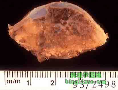 |
一例切下的有较宽基底的骨软骨瘤,但仍然由带软骨帽的多孔骨组成。 This is an example of an excised osteochondroma that has a broader base, but still consists of cancellous bone capped by cartilage. |
 |
骨软骨瘤的显微镜显示左边为良性软骨帽,右边为骨皮质。这种骨生长虽为良性,但有时会引起疼痛与刺激则需要外科切除。 The microscopic appearance of an osteochondroma displays the benign cartilagenous cap at the left and the bony cortex at the right. This bone growth, though benign, can sometimes cause problems of pain and irritation that leads to removal surgically. |
 |
骨巨细胞瘤。近端股骨已被截肢并切成两半而在干骺端显露出一暗红黑色血肿。骨巨细胞瘤放疗后可溶解。 Here is a giant cell tumor of bone. The proximal femur has been amputated and cut in half to reveal an irregular dark red-black hemorrhagic mass in the epiphyseal region. Giant cell tumors are lytic on radiography. |
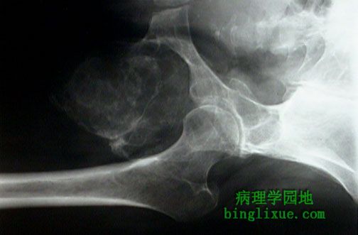 |
左坐骨支的骨巨细胞瘤X线图。肿瘤呈偏心性生长,可见膨胀的可溶性肿块,向反应性新生骨周围的软组织蔓延。 This is a radiograph of a giant cell tumor involving the left ischial ramus. The tumor is an eccentric, expansile, lytic mass with extension into soft tissue along with overlying reactive new bone formation. |
 |
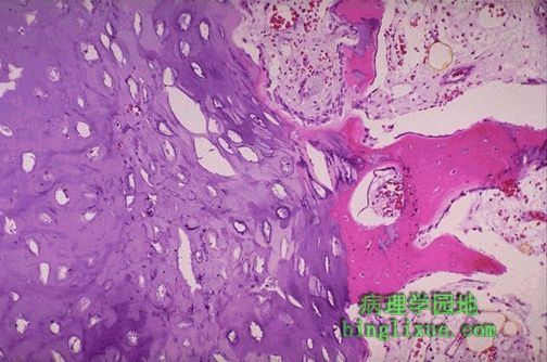
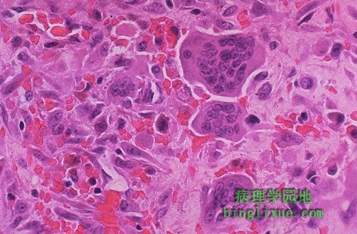
骨巨细胞瘤,是由大量圆形、卵圆形的单核基质细胞和分布于其间的多核巨细胞组成。生物学上这些肿瘤为良性且可通过刮除术或切除术治愈。如呈放射状,它们可能发生恶性转变。 Histologically, giant cell tumors of bone as seen here are composed of multinucleated giant cells in a sea of round to oval mononuclear cells. These tumors are biologically benign and are treated by curettage or resection. When radiated, they may undergo malignant transformation. |
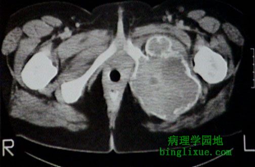 |
左坐骨支的骨巨细胞瘤CT图。肿瘤呈偏心性生长,可见膨胀的可溶性肿块,向反应性新生骨周围的软组织蔓延。 This is a computed tomographic (CT) scan of a giant cell tumor involving the left ischial ramus. The tumor is an eccentric, expansile, lytic mass with extension into soft tissue along with overlying reactive new bone formation. |
 |
股骨动脉瘤性骨囊肿的肉眼外观。受累骨通常为长骨干骺端或椎体脊,X线显示为典型的气囊化或动脉瘤样膨胀。大体上,围绕充满血的囊腔有着丰富的肿瘤聚集物。血管破坏出血形成的含血铁黄素使一些包囊染成褐色。 The gross appearance of an aneurysmal bone cyst is shown here. The radiographic appearance is typically that of ballooning, or aneurysmal dilation, of the affected bone--usually the metaphysis of a long bone or dorsal vertebral body. Grossly, there are fleshy aggregates of tumor surrounding cystic spaces filled with blood. The breakdown of the blood into hemosiderin has led to brownish staining of some of the cysts seen here. |
 |
低倍镜显示动脉瘤性骨囊肿。海绵状腔隙充满着血。腔壁包含梭形细胞与多核细胞。 The low power microscopic appearance of an aneurysmal bone cyst is seen here. Cavernous spaces are filled with blood. The walls of the spaces contain spindle cells and multinucleated cells. |
 |
高倍镜显示动脉瘤性骨囊肿。可见间质中突出的多核细胞与饱满的梭形细胞。 This is an aneurysmal bone cyst at high power magnification. Note the prominent multinucleated cells and the plump spindle cells of the stroma. |