
 |
脊柱矢状面MRI显示C7(第7颈椎)的转移瘤性病变,图上第一颈椎不能看到,颈5和颈6表现为先天性的部分融合。 This magnetic resonance imaging (MRI) scan of the spine in sagittal view demonstrates a metastatic lesion destroying C7 (the first cervical vertebra is not visible in this view, and there is partial congenital fusion of C5-C6). |
 |
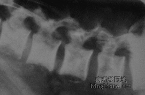
骨转移的较近视图。事实上所有骨的转移性病变都是癌。一般会转移到骨的原发癌的位置有:胸部、前列腺、肾、甲状腺、肺。肾细胞癌趋于导致骨质溶解(破坏骨)而前列腺癌趋于导致成骨(促使新骨形成)。 Here is a closer view of bone metastases. Virtually all bone metastases are from carcinomas. The primary sites of carcinomas that commonly go to bone are: breast, prostate, kidney, thyroid, lung. Renal cell carcinomas tend to be osteolytic (they destroy the bone) whereas prostatic adenocarcinomas tend to be osteoblastic (they initiate new bone formation). |
 |
前列腺癌病人X线显示脊椎转移性成骨性骨肿瘤。 This radiograph demonstrates osteoblastic metastases in the vertebral column in a patient with metastatic adenocarcinoma of prostate. Vertebral osteoblastic metastases |
 |
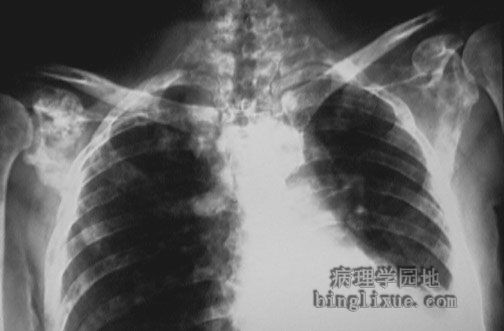
胸部X线片显示胸部转移性成骨性骨肿瘤。 This chest radiograph demonstrates osteoblastic metastases in a patient with metastatic adenocarcinoma of prostate. Chest, osteoblastic metastases |
 |
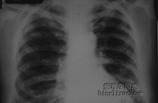
胸部X线显示,乳腺癌转移病人,胸部骨质溶解破坏。 This chest radiograph demonstrates osteolytic metastases in a patient with metastatic breast carcinoma. Chest, osteolytic metastases |
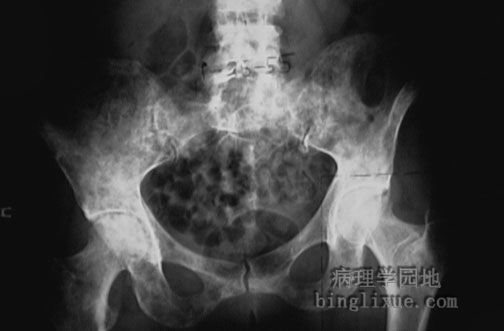 |
乳腺癌病人X线片显示转移性病变,盆部的转移性肿瘤破坏溶解骨质,为溶骨性转移性骨肿瘤。 This radiograph demonstrates osteolytic metastases in the pelvis in a patient with metastatic breast carcinoma. Pelvis, osteolytic metastases |
 |
转移性骨肿瘤显微镜视图。该区域的放射摄影扫描显现为“热点”。转移性病灶使骨明显脆弱,便可出现病理性骨折。 Here is a microscopic view of metastases to bone. Such areas appear as "hot spots" on radiographic scans. If the bone is markedly weakened by the metastasis, then a "pathologic" fracture is possible. |
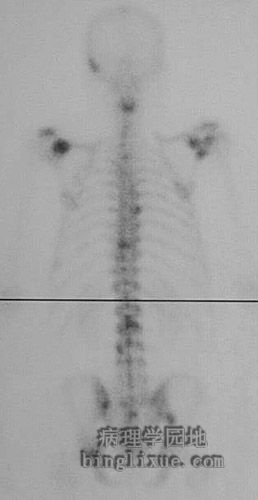 |
核素扫描显示多个病灶,脊柱周围颜色较深的病灶代表转移性病变。 This nuclear medicine bone scan reveals multiple areas of increased uptake, which are the darker foci, such as in the vertebral column representing metastases. |
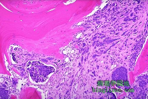 |
高倍放大显示转移的乳腺浸润性导管癌,骨内和骨髓腔内布满这种转移性病变。并有反应性的新骨生成,在靠近左上部骨板处呈现淡红色类骨质。 At high magnification, metastatic infiltrating ductal carcinoma of breast is seen within bone and filling the marrow cavity. There is reactive new bone with pale pink osteoid being laid down next to a bony spicule at the upper left. |
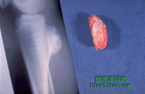 |
骨软骨瘤。病变呈现为骨性突起(外生骨疣)。多数为单发,局部疼痛时可能要切除。多发性骨软骨瘤罕见,其特征是骨畸形并有较大的发展成软骨肉瘤的倾向。 |