
 |
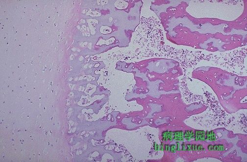
正常胎儿生长骨板,显示长骨的软骨内骨化过程。 This is the normal fetal growth plate demonstrating the process of endochondral bone formation in a long bone. |
 |
在新生骨的骨陷窝周可见成骨细胞排列,这些细胞在钙化的软骨化骨周围沉积成骨。 Osteoblasts can be seen lining the lacunae in the newly developing fetal bone, and they deposit bone along calcified cartilagenous spicules. |
 |
骨折区形成的不规则新生骨或称编织骨。不规则骨小梁周围可见成骨细胞排列,中心部位可见破骨细胞。 This is irregular new bone, or woven bone, which is forming in the region of a fracture. Osteoblasts are seen lining the irregular trabeculae, and there is an osteoclast near the center. |
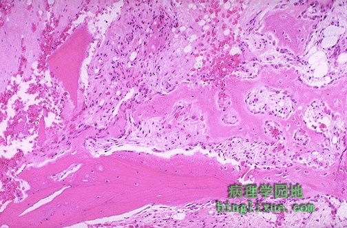 |
骨折区域的左侧和底部可见残存破损的骨小梁,右侧和顶部可见淡粉色的新生骨形成。 Here is a region of fracture with remaining disrupted trabeculae at the left and bottom. The paler pink new bone is forming at the right and top. |
 |
新近骨折区域,骨痂正在破碎的骨小梁末端形成,骨小梁从左上部扩展至中心处。 In this region of a recent fracture, callus is seen forming at the broken ends of bony trabeculae that extend to the center from the left and top. |
 |
慢性骨髓炎,可见骨髓腔的纤维化并伴有慢性炎细胞浸润。可能有重塑性骨破坏。骨髓炎很难治愈。 This is chronic osteomyelitis. Note the fibrosis of the marrow space accompanied by chronic inflammatory cells. There can be bone destruction with remodelling. Osteomyelitis is very difficult to treat. |
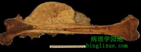 |
图示骨肉瘤,股骨骺端出现了一大半轮状肿块。多见于年轻人(可见右侧明显的骨骺)。 This femur has a large eccentric tumor mass arising in the metaphyseal region. This is an osteosarcoma (a variant known as parosteal osteogenic sarcoma) of bone. These tumors most often occur in young persons (note that the epiphysis seen at the right is still present). |
 |
此骨肉瘤来自一个十几岁的少年的胫骨上部干骺端,它穿越骨皮质延伸到软组织。肿瘤坚固呈灰白色。关节腔处可见闪亮的白色股骨髁的关节软骨恰好靠近肿瘤的右部。 This osteosarcoma arising in the metaphysis at the upper tibia of a teenage boy breaks through the bone cortex and extends into soft tissue. The tumor is firm and tan-white. The glistening white articular cartilage of the femoral condyle can be seen just to the right of the tumor in the opened joint space. |
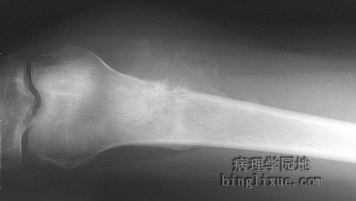 |
X线显示股骨远端干骺区的骨肉瘤。肿瘤侵袭破坏骨皮质,并向软组织蔓延,可见伴有钙化的不规则肿瘤性骨样组织。左侧可见骨膜被掀起。 This is a radiograph of an osteosarcoma involving the metaphyseal region of the distal femur. The tumor erodes and destroys the bone cortex, extending into soft tissue where irregular tumor bone with calcification is seen. At the left, the periosteum is being lifted off. |
 |
骨肉瘤的显微外观。肉瘤细胞有明显的多形性,常呈梭形。靠近中心处可见一很具有很大胞核的大肿瘤细胞。还可见反应性的新生骨岛。 The microscopic appearance of an osteosarcoma is shown here. Sarcomas have very pleomorphic cells, often with a spindle shape. One large cell with very large nuclei is seen near the center. There are islands of reactive new bone. |