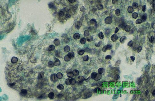

卡氏肺孢子虫肺炎:大体 大体放大 低倍镜 高倍镜 GMS染色
The best way to make the diagnosis of Pneumocystis carinii pneumonia is to perform a Gomori methenamine silver (GMS) stain on the lung tissue or bronchoalveolar lavage (BAL) fluid. The cyst wall is stained, and the organisms appear as crushed ping-pong balls, or crescent shapes, or folded spheres, or flattened beach balls, or deflated tennis balls, or....