
 |
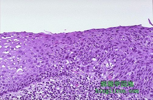
This is dysplasia. The normal squamous epithelium at the left transforms to a disorderly growth pattern at the right. This is farther down the road toward neoplasia. 图示:不典型增生 左侧的正常鳞状上皮转变为右侧的混乱排列模式。此为癌变的更危险的信号。 |
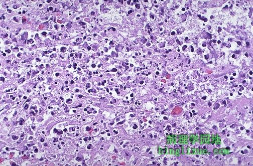 |
When there is marked cellular injury, there is cell death. This microscopic appearance of myocardium is a mess because so many cells have died that the tissue is not recognizable. Many nuclei have become pyknotic (shrunken and dark) and have then undergone karorrhexis (fragmentation) and karyolysis (dissolution). The cytoplasm and cell borders are not recognizable. 细胞损伤严重时,可导致坏死。光镜下显示心肌纤维紊乱,许多细胞坏死以后,使心肌组织不能辨认。 |
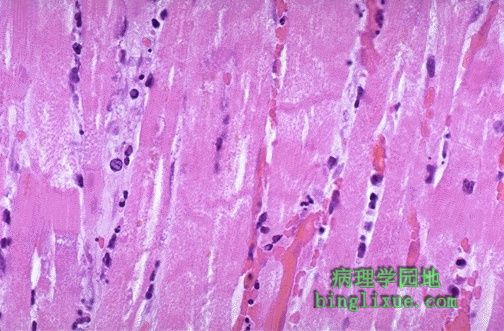 |
Here is myocardium in which the cells are dying. The nuclei of the myocardial fibers are being lost. The cytoplasm is losing its structure, because no well-defined cross-striations are seen. 图示:正坏死的心肌 心肌纤维的细胞核逐渐消失,细胞质结构破坏,横纹变得模糊不清。 |
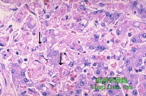 |
Apoptosis is a more orderly process of cell death in which there is individual cell necrosis, not necrosis of large numbers of cells. In this example, liver cells are dying individually (arrows) from injury by viral hepatitis. The cells are pink and without nuclei. 细胞凋亡是一种程序性细胞死亡,属于单个细胞死亡,而不是大量细胞死亡。图为肝细胞调亡,箭头所指为由肝炎病毒所致的凋亡细胞。呈粉红色,无细胞核。 |
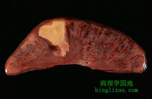 |
When many cells undergo necrosis at once, then definable patterns of necrosis are produced, depending upon the nature of the injury, the type of tissue, and the length of time. This is an example of coagulative necrosis. This is the typical pattern with ischemia and infarction (loss of blood supply and resultant tissue anoxia). Here, there is a wedge-shaped pale area of coagulative necrosis (infarction) in the renal cortex of the kidney. 大量细胞死亡即为坏死。坏死的类型取决于损伤的性质、组织的类型和持续的时间长短。图为凝固性坏死。是由缺血引起的典型坏死类型。图示:肾脏肾皮质区的楔型苍白区域即为凝固性坏死(梗死)。 |
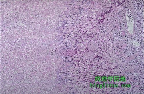 |
Microscopically, the renal cortex has undergone anoxic injury at the left so that the cells appear pale and ghost-like. There is a hemorrhagic zone in the middle where the cells are dying or have not quite died, and then normal renal parenchyma at the far right. This is an example of coagulative necrosis. 图示:凝固性坏死 镜下观:左侧的肾皮质遭受了缺氧性损伤后,细胞呈栅栏状排列。中间是细胞未完全坏死的出血带。最右侧是正常的肾实质。 |
 |
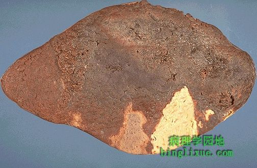
The contrast between normal adrenal cortex and the small pale infarct is good. The area just under the capsule is spared because of blood supply from capsular arterial branches. This is an odd place for an infarct, but it illustrates the shape and appearance of an ischemic (pale) infarct well. 正常的肾上腺皮质和梗死的苍白色小斑块间的界限是分明的。被膜下的坏死区域恰在相应动脉分支的供血区。这虽然是单个梗死灶,但它说明了梗死灶的形状和缺血区的关系。 |
 |
Two large infarctions (areas of coagulative necrosis) are seen in this sectioned spleen. Since the etiology of coagulative necrosis is usually vascular with loss of blood supply, the infarct occurs in a vascular distribution. Thus, infarcts are often wedge-shaped with a base on the organ capsule. 图示:脾切面的两个大梗死灶(凝固性坏死)。梗死的原因通常是供血不足,因此梗死灶位于相应动脉分支的供血区域。因此梗死灶的形状常为底朝向器官被膜的楔形。 |
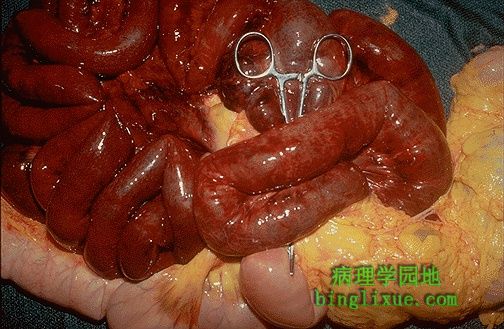 |
The small intestine is infarcted. The dark red to grey infarcted bowel contrasts with the pale pink normal bowel at the bottom. Some organs such as bowel with anastomosing blood supplies, or liver with a dual blood suppy, are hard to infarct. This bowel was caught in a hernia and the mesenteric blood supply was constricted by the small opening to the hernia sac. 图示:小肠出血性梗死(又称红色梗死)。暗红甚至是灰色的梗死肠段与下部的粉红色正常肠段有着明显的区别。一些器官如肠侧支循环较多,或者如肝有双重血液供应,很难发生梗死。但这段肠发生了肠疝,由于疝环很小,因此肠系膜动脉供血明显减少。 |
 |
The two lung abscesses seen here are examples of liquefactive necrosis in which there is a liquid center in an area of tissue injury. One abscess appears in the upper lobe and one in the lower lobe. Liquefactive necrosis is typical of organs in which the tissues have a lot of lipid (such as brain) or when there is an abscess with lots of acute inflammatory cells whose release of proteolytic enzymes destroys the surrounding tissues. 图示:肺的两个液化性坏死灶。在损伤组织区域中央发生液化。一个脓肿在肺上叶,一个在肺下叶。液化性坏死是含有较多脂质器官(如脑)的坏死类型。当大量的急性炎症细胞释放蛋白溶解酶破坏周围组织时,即发生脓肿(液化性坏死)。 |