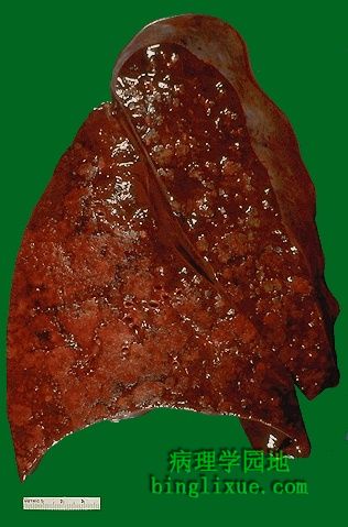
肺切面显示典型的灰黄*色实变区域的支气管肺炎。由于明显的肺充血,残余的肺组织呈暗红色。支气管肺炎(小叶性肺炎)出现典型的不规则实变病灶,病变以肺小叶为中心。图片左下部左下肺叶显示多病灶融合。实变区域比周围的肺组织坚韧。
The cut surface of this lung demonstrates the typical appearance of a bronchopneumonia with areas of tan-yellow consolidation. Remaining lung is dark red because of marked pulmonary congestion. Bronchopneumonia (lobular pneumonia) is characterized by patchy areas of pulmonary consolidation. These areas become almost confluent in the left lower lobe on the bottom left of the photograph.The areas of consolidation are firmer than the surrounding lung.

