
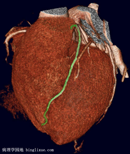
血管造影后多层螺旋CT显示冠状面的冠状动脉主干。绿色线条为左冠状动脉前降支,从图上不易看出左前降支源于左冠状动脉主干。近端几粒米的LAD(左冠状动脉前降支)表现出不规则的狭窄,是由冠状动脉粥样硬化所导致的。虽然生活方式的改变能够逆转动脉粥样性狭窄,但是急性冠脉事件出现时,就需要考虑冠脉搭桥术或冠脉成形术,当然也需要考虑其它主干的受累情况。血管造影后多层螺旋CT显示技术对近心外膜的冠状动脉狭窄具有很好的敏感性和特异性,可以替代血管造影后冠状动脉插管术。
This is once slice of a multi-slice CT (MSCT) angiogram demonstrating the major coronary arteries of the heart in coronal view. In this view that highlights the left anterior descending artery (in green) it is difficult to clearly outline the origin from the left main coronary, or tell the relationship to the left circumflex. However, there is irregular narrowing of the LAD proximally for the first few centimeters, consistent with atherosclerotic coronary artery disease. Such atherosclerotic narrowing may be amenable to lifestyle changes to reverse the atherosclerosis, but if an acute coronary syndrome has occurred, then bypass grafting or angioplasty may be considered, with consideration of the extent of other major coronary vessel involvement. Coronary angiography by MSCT has good sensitivity and specificity for detecting significant narrowing in proximal epicardial coronary arteries, and can replace cardiac catheterization with angiography for diagnostic purposes.