
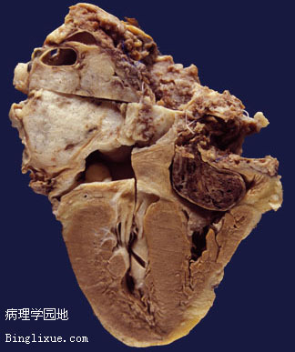
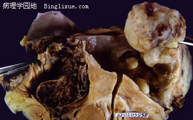
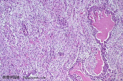
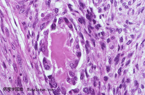
55岁女性病人,图示一累及左心房的肿块性病变,右心房见血栓。肿块呈白色到深棕色、形状不规则,侵蚀附近组织。显微镜下见深染多形性的梭形或椭圆形细胞。局部可见粉红色类骨质。显微镜下见转移到肺。原发性心脏骨肉瘤罕见,只占心脏肿瘤的1%,通常源于左心房。钙化可能存在。 转移性骨肉瘤通常累及右心。原发性心脏肿瘤可以产生许多其他心脏病的表现,可能阻塞血流,引起心力衰竭、胸痛、晕厥、肺动脉高压和心律失常等。 CT扫描肿能有效帮助确定肿块的位置和结构。
There is a mass lesion involving the left atrium, with a thrombus in the right atrium, from a 55 year old woman. The mass is white to tan and irregular, invading adjacent structures. Microscopically, there are spindle to oblong cells with hyperchromatism and pleomorphism. Focally, pink osteoid is present. There were microscopic metastases to the lung. Primary cardiac osteosarcomas are rare, accounting for only 1% of all cardiac tumors, and they usually originate in the left atrium. Calcifications may be present. Mestatatic osteosarcomas usually involve the right side of the heart. Primary cardiac tumors can mimic many other cardiac conditions and may cause obstruction to blood flow, heart failure, chest pain, syncope, pulmonary hypertension, and arrhythmias. CT scanning may be useful to determine the location and composition of a cardiac mass.