
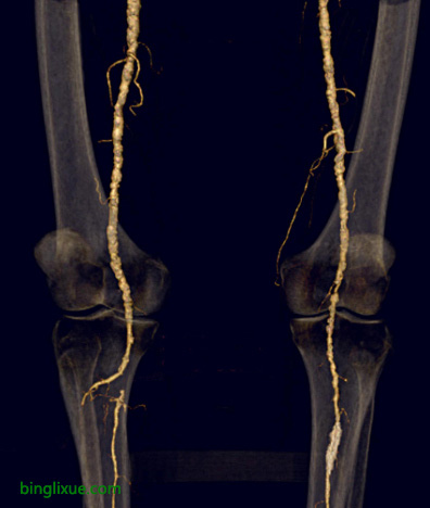
动脉粥样硬化可能涉及外周动脉,特别是腿部,从髂动脉开始并延伸至股动脉,腘动脉和小腿动脉。图示CT血管造影,右侧腘动脉闭塞,可见有血流进入侧支动脉分支。并发症包括运动耐量降低、疼痛和缺血性改变,最终导致坏疽。
踝肱指数(ABI)可检测外周血管疾病,可用袖带和多普勒超声测量。首先在手臂中部的肱动脉水平测量收缩压,然后在踝的水平测量收缩压。 ABI是踝部收缩压与肱动脉收缩压的比值。 ABI通常应为1.0。低于0.85可诊断为外周血管疾病。
另一种测量是动脉血液流入腿部时发生的体积变化的容量记录(PVR)。通过沿着大腿、小腿、脚踝和大脚趾放置多个压力袖带来执行PVR。袖带充气至40-60mmHg。腿中的体积变化与通过换能器记录的袖带中的压力随时间的变化相关。
Atherosclerosis may involve peripheral arteries, particularly the leg, starting from the iliac arteries and extending to femoral, popliteal, and lower leg arteries. Shown here with CT angiography is an occlusion of the right popliteal artery. There is runoff into a collateral arterial branch. Complications include reduced exercise tolerance, pain, and ischemic changes that can culminate in gangrenous necrosis.
The ankle-branchial index (ABI) can document peripheral vascular disease and is measured with a manual blood pressure (BP) cuff and a Doppler ultrasound device. With DU the systolic arterial occlusion pressure is measured, first at the level of the brachial artery in the arm, then at the level of the ankle. ABI is the ratio of ankle occlusion pressure to brachial artery occlusion pressure. The ABI should normally be 1.0. An ABI below 0.85 is consistent with PAD.
Another measure of peripheral vascular disease that performed along with ABI is pulse-volume recording (PVR) of volume changes that occur with arterial blood flow into the leg. PVR is performed by placing multiple pressure cuffs along the thigh, calf, ankle, and great toes. The cuffs are inflated to 40-60 mm Hg. Changes in the volume in the leg correlate with changes in pressure in the cuff that are recorded via a transducer over time.